POSITION
Copyright 2007
Ascending Aorta
Annulus
The annulus is located at the base of the aorta and represents the junction between the aorta and the left ventricle. It also is connectes to the annulus of the mtral valve both lying at the crux of the heart at the intersection of the atrial septum and ventricular septum and conal septum.
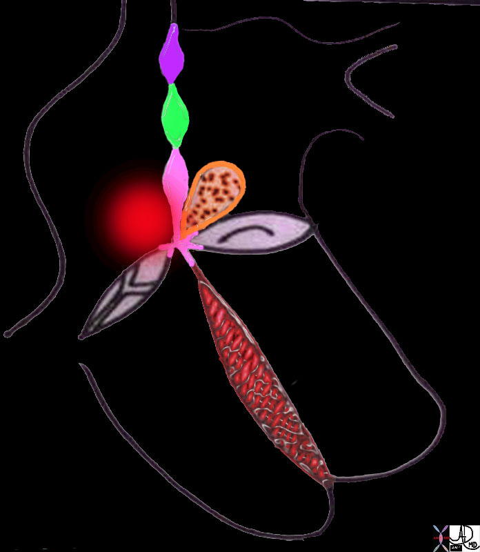
Aortic Annulus is attached to the Crux of the Heart |
| 01669b04 heart cardiac embryology conal septum line drawing right atrium left atrium left ventricle right ventricle interatrial septum interventricular septum atrioventricular septum crux of the heart embryology anatomy Davidoff drawing Davidoff MD 01667b04 01667b04 01667b06 01667b07 01667b09 01667b14 01669b03 |

Aortic Valve |
| The aortic annulus red) and valve (AV) lies central to many structures including the right ventricular outflow tract (RVOT), pulmonary valve(PV), left atrium (LA), right atrium (RA), and superior |
Aortic Valve
The aortic valve is aligned along the rightward axis of the aorta. The complex of structures including the annulus, valve, sinus and and proximal portion of the ascending laorta occupy the most comfotrtable position of the body. They lie between the two low pressure parts of the heart, the right and left atriumie between the annulus and the aortic valve on the upstream side, and the tubular portion of the ascending aorta on the other side.
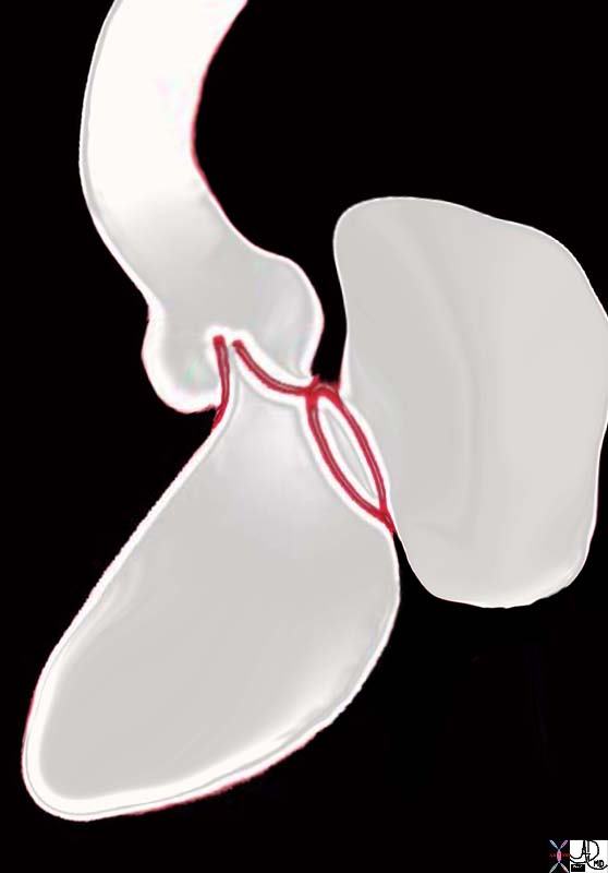
Aortic Valve in Fibrous Continuity with the Mitral Valve |
| 06373b05 heart cardiac aorta mitral valve anatomy embryology resorbtion of subaortic conus fibrous continuity mitral valve with aortic valve position shape of aorta MV Courtesy Ashley Davidoff Davidoff drawing |
Sinuses
The Sinuses are aligned along the rightward axis of the aorta and lie between the annulus and the aortic valve on the upstream side, and the tubular portion of the ascending aorta on the other side.
Tubular Portion
Aortic Arch
The arch measures about 2.5-3cms
Isthmus
At the isthmus the aorta narrows by about 3mm. The isthmus defines the attachment of the ligamentum arteriosum to the aorta as well as delineating the arch from the descending aorta.
Abdominal Aorta
ding

Sagittal position within the body |
| 49168 normal aorta sagittal Courtesy Ashley Davidoff MD Uploaded RP |
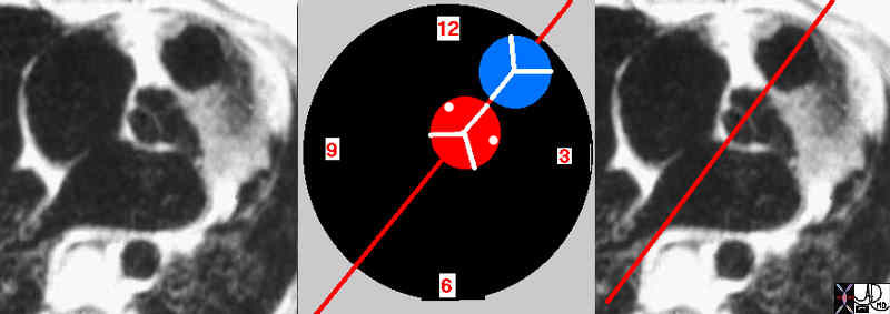
Position of the aorta relative to the subpulmonary conus |
| 07954eW.800 aorta aortic valve infundibulum RVOT right ventricular outflow tract position relation MRIscan Davidoff MD |
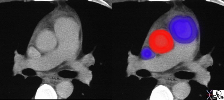
Aorta and Pulmonary Artery – Normal Size and Position |
| 27467c01 aorta pulmonary valve pulmonary artery SVC size position normal CTscan Davidoff MD |
The abdominal aorta begins at the median, aortic hiatus of the diaphragm, anterior to the twelfth thoracic vertebra. (1) It descends anterior to the vertebrae to end at the fourth lumbar, slightly to the left of midline, and divides into two common iliac arteries.
Bicuspid Aortic Valve and Malposition of the Coronary Ostium
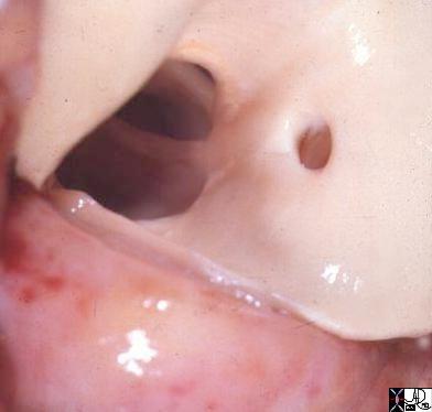
Bicuspid Aortic Valve and Malposition of Coronary Ostium |
| 15049 aorta aortic valve bicuspid aortic valve hypoplastic valve anomalous positioning of a coronary ostium coronary artery congenital abnormality position gosspathology Davidoff MD |
D Transposition of the Great Vessels
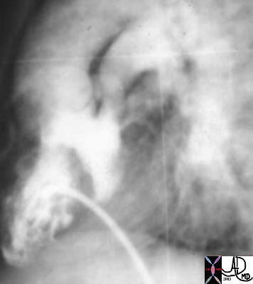
Transposition of the Great Vessels |
| This is an angiogram of the RV, showing an anteriorly placed aorta, a VSD filling the LV, and a posteriorly positioned smaller MPA. The catheter courses via the IVC into the RV. The findings are consistent with TGA and in this case a D-TGA, though it is impossible to identify the position of the aortic valve in relation to the PA in this lateral projection. An associated VSD and subpulmonary stenosis and or PS is implied by the small sized PA. Courtesy Ashley Davidoff MD 01487 code CVS heart cardiac transpoistion of the great arteries DTGA VSD subpulmonary stenosis imaging radiology angiography |
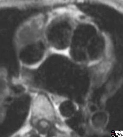
DTGA |
| 07258b02 anterior aorta posterior pulmonary artery position connection subaortic conus D TGA TGV D transposition of the great vessels D transposition of the great arteries dextro rightward MRI Davidoff MD |
Corrected Transposition
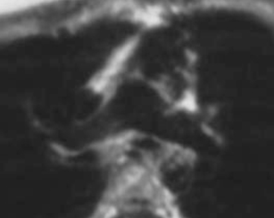
LTGA |
| 07304b01 anterior aorta posterior pulmonary artery position connection subaortic conus L TGA L TGV L transposition of the great vessels L transposition of the great arteries levo leftward MRI Davidoff MD |
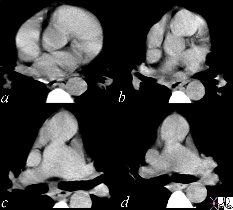
LTGA |
| 28999b01 heart cardiac aorta pulmonary artery RVOT conotruncal malformation LTGA L TGV transposition of the great vessels transposition of the geat arteries corrected transposition position connection relation embryology CTscan Davidoff MD 28994 28995 28996 28997 28998 28999 |
Right Aortic Arch

Dwarf with Kyphosis and secondary hypertrphy of the rectus abdominis muscles |
| 49838c05 50 year old female with respiratory difficulty trachea bronchi rectus abdominis muscle compression fractures kyphosis dwarf dwarfism right aortic arch tracheomalacia tracheal stenosis rectus abdominis muscle hypertrophy Davidoff MD Courtesy Ashley Davidoff MD CTscan 49838 49838c01 49838c02 49838c03 49838c04 49838c05 shape size position character growth |
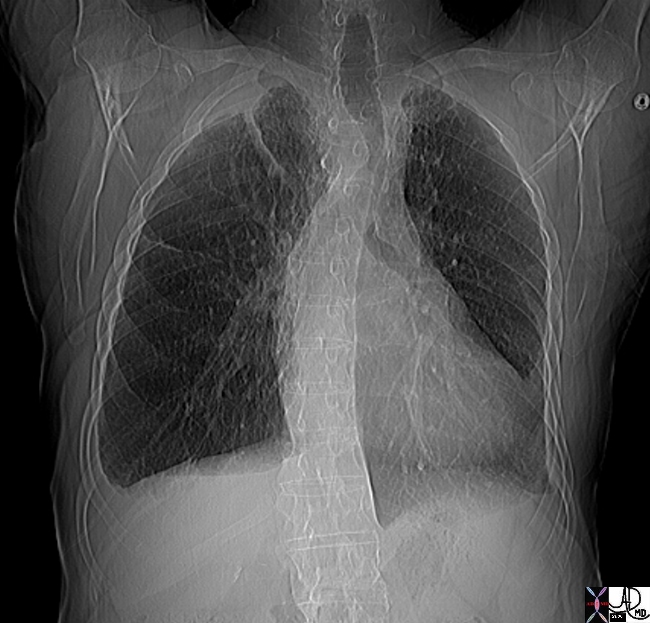
Righ Sided Impression on the Trachea |
| 76032 aorta arch right aortic arch left aortic arch normal position trachea impression on right side CTscan CourtesyAshley Davidoff MD |
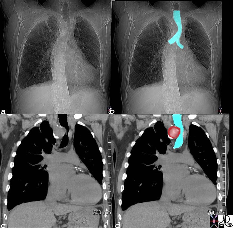
Normal and Right Aortic Arch |
| 76034c05 aorta arch right aortic arch left aortic arch normal position Ctscan CourtesyAshley Davidoff MD |
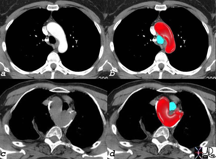
Normal and Right Aortic Arch |
| 76034c05 aorta arch right aortic arch left aortic arch normal position Ctscan CourtesyAshley Davidoff MD |
