
The Common Vein Copyright 2012
Aortic Dissections
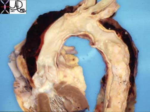
This pathological specimen shows an aortic dissection starting at the root of the aorta and extending across the arch and into the descending portion. The false lumen is filled with clotted blood.
Courtesy of: Henri Cuenoud, M.D.
Aortic dissection is a sudden catastrophic disruption of the aortic wall caused by a shearing tear of the intima, and secondary involvement of the media by the advancing dissection and hematoma. The event characterized by a splitting of the arterial wall along the longitudinal plane of the aorta.
The pain of aortic dissection is dramatic. It is classically hyper acute, sudden, severe, lancinating, excruciating, tearing, stabbing, throbbing, reaching its maximum intensity at the moment of onset. On the other hand, some patients may present with minimal or no symptoms. The duration of the pain is not measured in minutes and may simulate the pain of myocardial infarction. The pain may be felt in the anterior chest, the neck, and/or the back, sometimes reflecting the location of the dissection with ascending dissections noted with anterior chest pain, arch dissections with neck pain and descending thoracic dissections noted with intrascapular or back pain. Since dissections may involve the coronary arteries, and more commonly the right, myocardial ischemic pain may be present as well.
It is historically interesting to note that in 1910 William Osler recognized that “spontaneous tear of the arterial coats is associated with atrocious pain, with symptoms, indeed, in the case of the aorta of angina pectoris and many instances have been mistaken for it”.
The pain is caused by the stretching of the aortic wall, and hence its potential to have a throbbing character. It is not aggravated by position or body movement, nor relieved other than by analgesia, tincture of time, or repair.
The impending sense of doom originally described as “angor animi” literally means anguish of the soul may accompany the pain. This sensation sometimes accompanies other life-threatening situations when there is massive outpouring of catecholamines, such as myocardial infarction and pulmonary embolism.
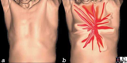
AcuteAorticSyn.jpg
Acute Severe Lancinating, Shearing, Chest Pain of Acute Aortic Syndrome
Ashley Davidoff
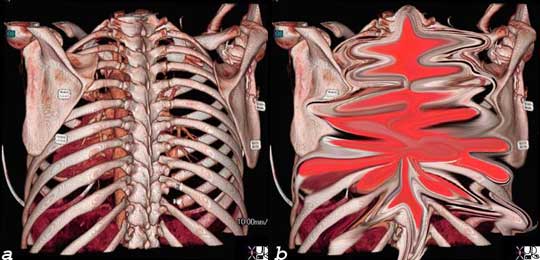
The pain of dissection may be felt either in the front of the chest and/or the back. Either way, the pain can have similar characteristics – i.e. sharp, thunderbolt-like, severe, and rapidly and almost instantaneously reaching maximum intensity coincident with the onset.
Courtesy of: Ashley Davidoff, M.D.
- spontaneous splitting the media of the aortic wall
- longitudinal separation of the aortic intima and adventitia
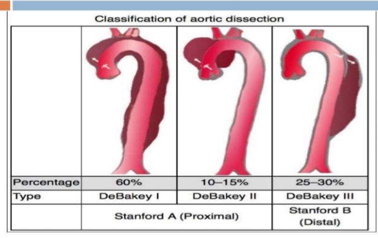
Stanford type A dissection 60%–70% of cases
- involves the ascending thoracic aorta,
- dissection flap may extend into the descending aorta.
- untreated, type A dissections are associated with a mortality rate of over 50% within 48 hours
Fatal Acute Dissection
This pathological specimen shows an aortic dissection starting at the root of the aorta and extending across the arch and into the descending portion. The false lumen is filled with clotted blood.
Courtesy of: Henri Cuenoud, M.D.
Dissection of the Ascending Aorta
Ashley Davidoff
TheCommonVein.net
CXR
- normal in 10%–40%
- displacement of calcification of the aorta was reported in 15%
- abnormal cardiac contour being noted in 25%
- Waterbottle heart
- Transthoracic echocardiography
- Type A
- sensitivity 80% -100%
- Type B
- 30%–50% for dissections involving the descending aorta
- Type A
- TEE
- can be performed in the ER
- requires expertise
- 95%–100% specificity
- distal ascending aorta
- brachiocephalics and carotids
- not be adequately evaluated
- good for
- aortic root dilatation,
- aortic regurgitation,
- coronary ostial patency,
- pericardial effusions,

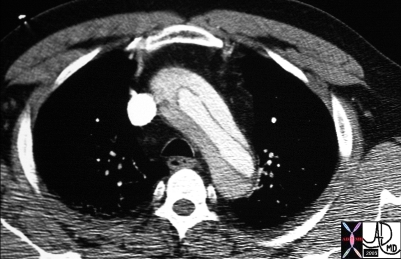
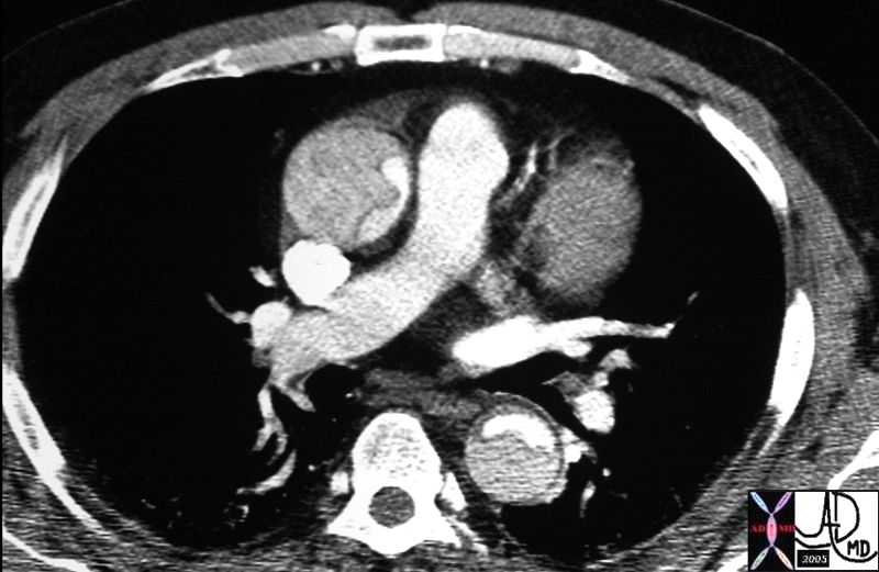
The CT image shows a dissection in the ascending aorta. The lumen consists of the smaller true lumen (with white contrast) and the false lumen (larger with grayer density). The involvement of the ascending aorta makes it a type A dissection and therefore, treatment requires surgical repair. In this patient, however, the dissection extended to the descending aorta as well. Either way a type A dissection requires surgery.
Courtesy of: Ashley Davidoff, M.D.
Type A Aortic Dissection |
|
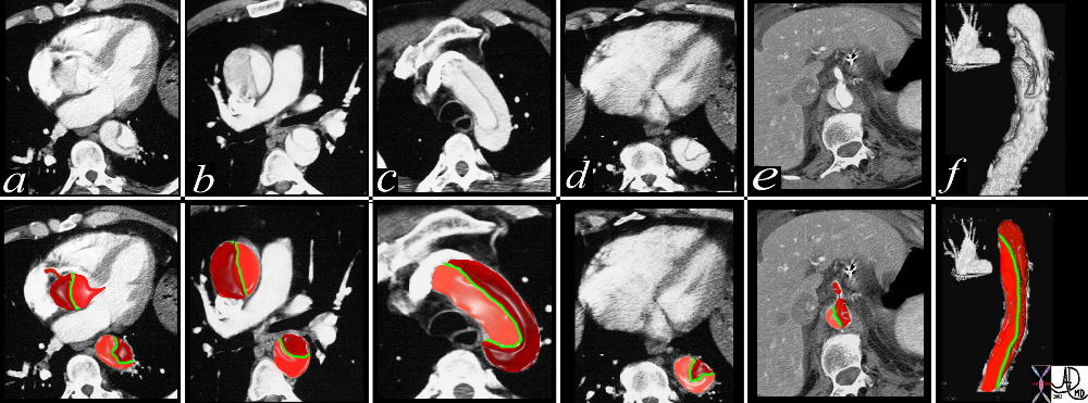
This combination of images from a CTscan through the thorax, reveals an aortic dissection starting at the level of the aortic valve and extending into the abdomen with involvement of the celiac axis. The true lumen is overlaid in pink while the false lumen is overlaid in a darker red. The green structure is the intimal flap. Flow in the false lumen is slightly delayed and slower as seen by the lower density of the contrasyt within it. Copyright 2012 Courtesy Ashley Davidoff MD. 19412c02 code CVS artery AO aorta dissection fx crescent |
Arch Dissection – Narrowed True Lumen |
|
thorax thoracic aorta arch aorta fx dissection dx aortic dissection type A CTscan Copyright 2012 Courtesy Ashley Davidoff MD 20449 |
36865c |
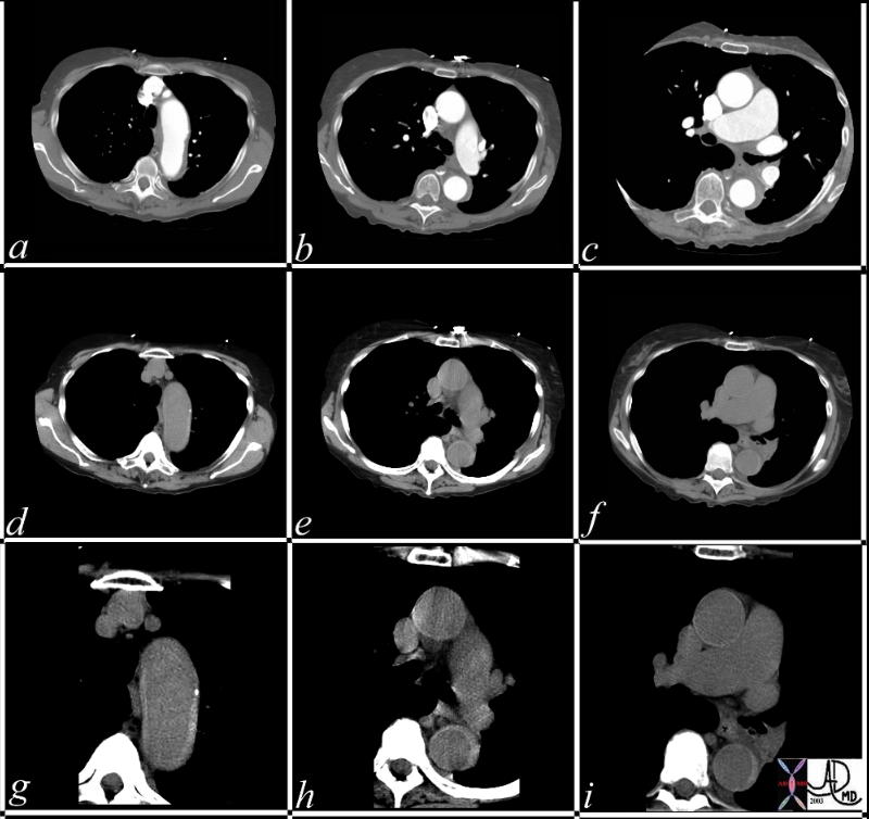
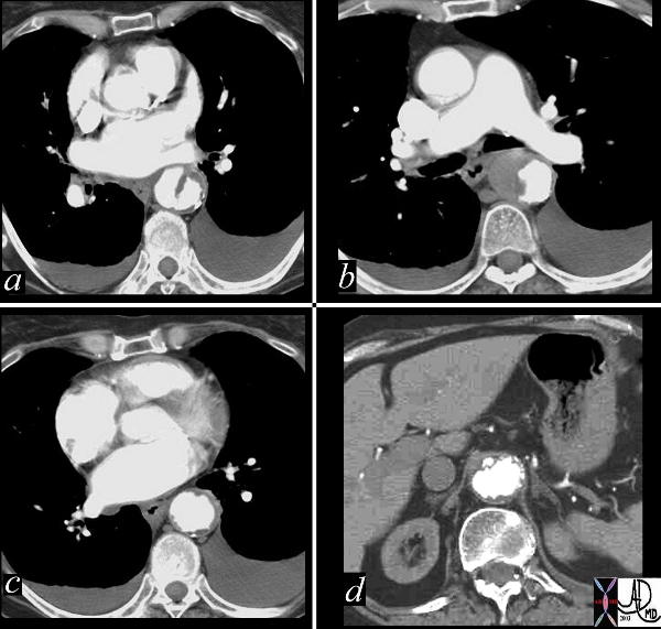
| This series of CT scans were taken 1 day apart. The patient presented with chest pain and the soft tissue changes around contrast filled aorta (a,b,c) suggested a chronic dissection, or an acute thrombosed dissection. 1 day later the non-contrast CT clearly reveals an acute thrombosed dissection (d,e,f) that started in the arch and extended along the descending thoracic aorta. The last series (g,h,i) enhance the non contrats study. A non contrast CT followed by contrast injection, is an important tchnique to optimally characterise acute changes in this clincal setting.
Copyright 2012 Courtesy Ashley Davidoff MD 36865c |
- Stanford type B dissection
- involves the descending thoracic aorta
- distal to the left subclavian artery
- 30%–40% of cases
- medical treatment of hypertension,
- unless complications
- extension of the dissection –
- end-organ ischemia
- persistent pain
- requires surgery
- extension of the dissection –
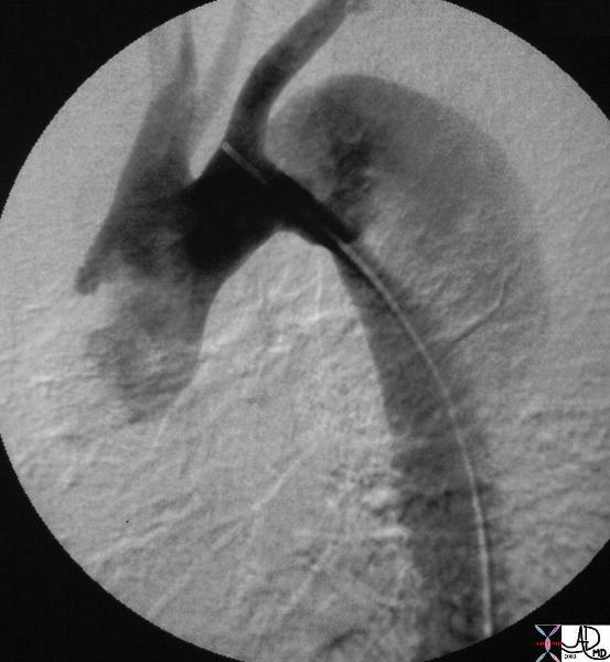
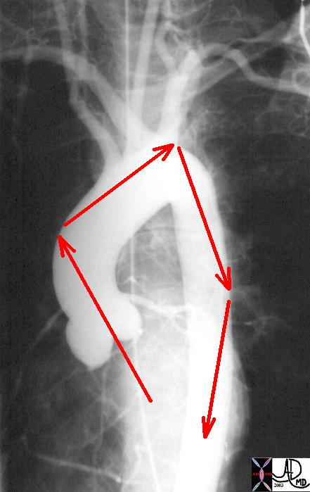
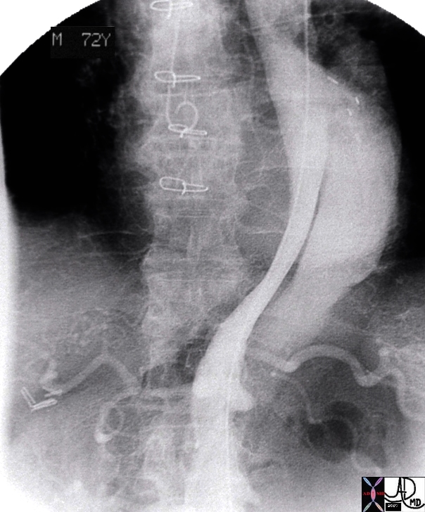
The digital angiogram in the LAO projection shows an intimal dissection starting just after the left subclavian artery. The true lumen is medial and smaller than the laterally and leftward placed larger false lumen. 35204 Courtesy Laura Feldman MD. code CVS artery aorta thorax thoracic descending dissection angiogram code aorta artery dissection flap.
Courtesy of Laura Feldman MD. 35204
Thoracic Aorta – Dissection |
|
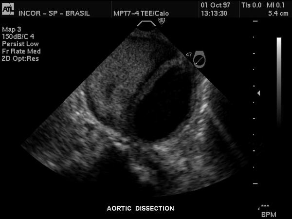
Gray scale US of the descending thoracic aorta showing a hypoechoic true lumen (black) and homogeneous echoes of the thrombosed lumen in this patient with aortic dissection. Courtesy Philips Medical Systems 33167 |
Color Flow Doppler – Descending Thoracic Aorta Dissection |
|
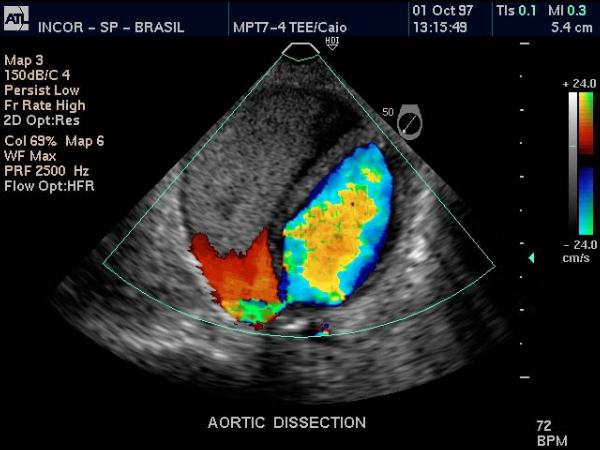
Doppler US of the descending thoracic aorta showing flow in the true lumen (color) and no flow in the thrombosed lumen (gray echoes) in this patient with aortic dissection. Courtesy Philips Medical Systems 33166 |
Atelectasis Masquerading as a Dissection |
|
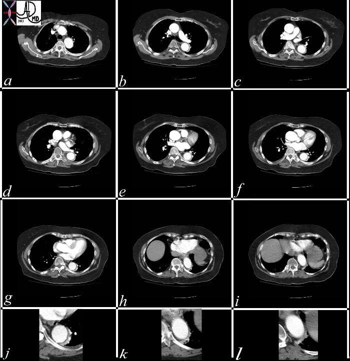
This series of CT scans from the arch to the abdominal inlet shows a curvilinear density around the aorta on its left lateral edge that masquerades as a dissection The last three images (g,h,i) and their magnified counterparts below them (j,k,l) shows air bronchograms opacified vessels within the crescentic density, and deviation from the contour of the aorta, confirming the atelectatic nature of the crescent. Copyright 2012 Courtesy Ashley Davidoff MD 37071c |
Dissection Caused by Ulcerating Plaque Complicated by Mural Hematoma |
|
This combination of images reflects the CTscan of a patient who presented with acute chest pain, and has findings reflecting a focal dissection of an atheromatous aorta. Note in (a) how thick the “intimal” flap is. Note also the mural dissection or hematoma with no flow within it. (b) Other parts of the descending aorta (c) and abdominal aorta (d) show only severe atheromatous disease. Copyright 2012 Courtesy Ashley Davidoff MD. 19416c |
True Lumen Compressed and Supplying Right Renal Artery |
|
artery aorta fx dissection dx dissection angiography Copyright 2012 Courtesy Ashley Davidoff MD 17474 |
True Lumen Compressed on Left Slow Flow in False Lumen |
|
abdomen artery + abdominal aorta + fx crescentic crescent shaped false lumen fx dissection + dx iatrogenic + imaging radiology CTscan radiologists and detectives Copyright 2012 Courtesy Ashley DAvidoff MD 19408 |
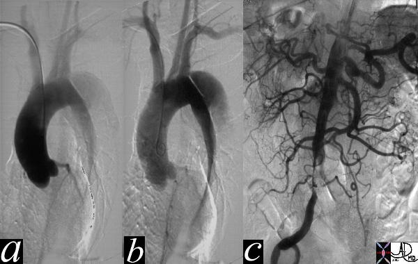
Type B Dissection |
|
The thoracic angiogram reveals an aortic dissection which starts just after the left subclaian artery. The true lumen is the narrowed medial lumen which is denser than the larger lateral false lumen. (a,b) In (c) the A-P view reveals a patent proximal abdominal aorta, but the distal narrowing and occlusion of the the left iliac artery suggests that the dissection has progressed distally. In the image 35124c the cross sectional images of the abdominal aorta reveal that the true lumen of the distal aorta is in fact quite narrowed. Courtesy Laura Feldman MD. 35116c |
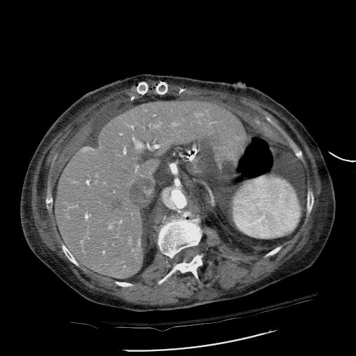
Chronic Aortic Dissection |
|
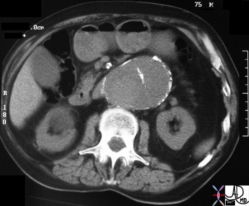
hx 75 male with abdominal pain abdomren abdominal AAA aneurysm fx mural calcification thrombosed aortic dissection chronic dissection CTscan Copyright 2012 Courtesy Ashley Davidoff MD 31262 |
Acute Traumatic Dissection |
|
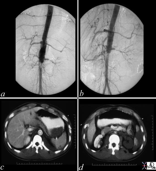
These images represent a traumatic dissection of the abdominal aorta caused by a compression injury to the abdomen. The angiograms in a and b shows what appears to be a complex tear of the aorta with a dissection and then a subtotal obstruction of the aorta distally. The CTscan shows (c) thedissection to better effect, with a subtotal narrowing of the aorta distally. Not also the injury of the left renal artery on the angiogram as well as the lack of perfusion of the left kidney on the CT scan. Copyright 2012 Courtesy Ashley Davidoff MD. 14734c |
Clinical
The clinical presentation is usually dramatic with hyperacute “shearing” or “ripping “chest or back pain. Proximal Anterior chest pain is usually associated with tears of the ascending aorta, while dissection of the descending aorta usually gives interscapular pain. Most patients are hypertensive. Findings on examination include aortic insufficiency (50%), pulse deficits (50% – commonly involve the brachiocephalic vessels) or neurologic manifestations Distal dissections may reveal pulse deficits of the left subclavian or femoral vessels
Diagnostic Tools
The radiologic diagnosis is often suspected on the plain film with the widened mediastinum and more specifically a blunted aortic knob or thickening of the wall of the knob more than 5 mm past the calcified aortic intima (Ring sign). CT, MRI and trans-esophageal echocardiography are all used with high sensitivity and specificity. TEE sensitivity/specificity of 99/98% Aortic angiography is an invasive procedure but may be best at delineating the extent of dissection
Dissection Involving the Ascending and Descending Aorta |
|
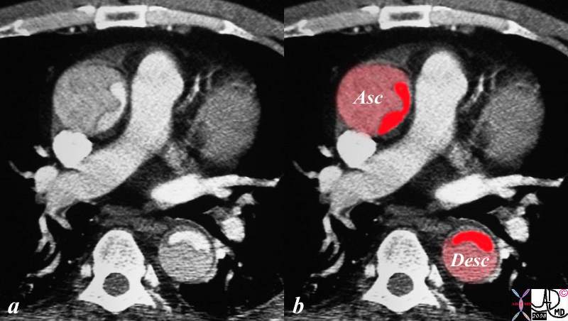
descending aorta ascending aorta fx dissection fx crescent dx aortic dissection type A CTscan Copyright 2012 Courtesy Ashley Davidoff MD 20448 |
Dissection Involving the Ascending and Descending Aorta |
|
The CT image shows a dissection in the ascending aorta. The lumen consists of the smaller true lumen (with white contrast) and the false lumen (larger with grayer density) The patient presented with classsical sever chest pain that radiated to the back. The involvement of the ascending aorta make s it a type A dissection and therefore treatment requires surgical repair. In this patient however the dissection extended to the descending aorta as well. Either way a type A dissection requires surgery. Copyright 2012 Courtesy Ashley Davidoff MD 20448c03.8s |
Type B dissection involving descending thoracic and abdominal aorta |
|
thorax abdomen fx dissection true lumen false lumen occluded renal artery angiogram angiography Copyright 2012 Courtesy Ashley Davidoff MD 24588b01 |
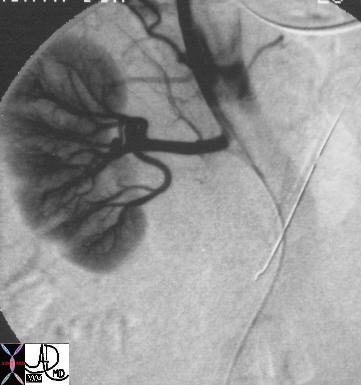
Hepatic Artery Dissection
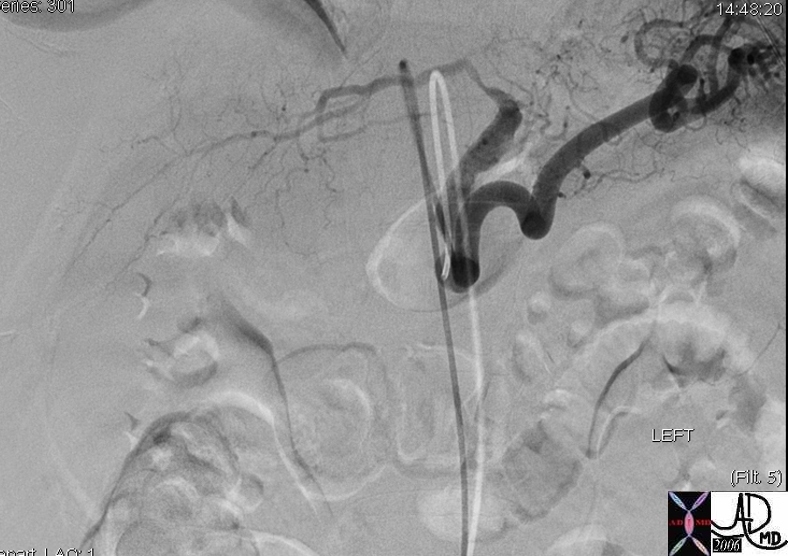
50 year old male presents with abdominal pain to the ER liver left hepatic artery fx focal aneurysmal dilatation of the proximal left hepatic artery fx focal narrowing of the mid portion at site of dissection dx spontaneous dissection of the left hepatic artery complicated by intraperitoneal rupture., treated by coil embolisation The common and right hepatic artery arose from the SMA
Copyright 2012 Courtesy Ashley Davidoff MD 45278b
Links and References
McMahon M et al Review of Aortic Dissection Radiographics 2010
Murillo H et al Aortic Dissection and other Acute Aortic Syndromes Radiographics 2021