
Character
Copyright 2007
The aortic wall, with its high elastic content supported by muscle and collagen, allows for transmission of a more or less steady blood pressure within the circulation by providing a tensile strength that can be adjusted to conform to the pumping strength of the heart muscle
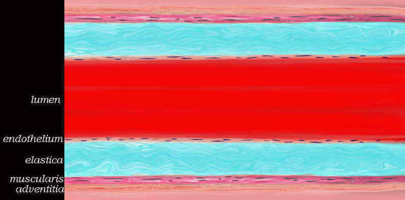
keywords artery aorta histology character normal endothelium media adventitia tunica elastica tunica muscularis.
Ashley Davidoff TheCommnonVein.net 47678b04.800
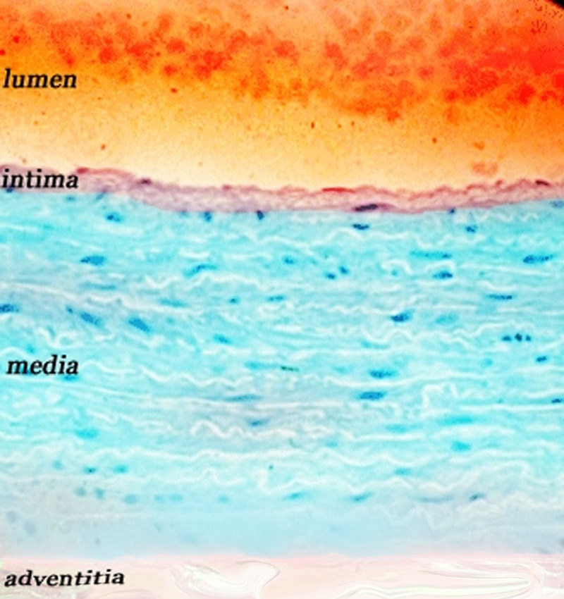
Histologic section of the wall of the aorta shows the thin intima, the thick elastic media characterised by the white wavy bands, and the media which consists of strong collagenous fibers.
keywords artery aorta histology character normal endothelium media adventitia tunica elastica tunica muscularis
Davidoff art Ashley Davidoff TheCommnonVein.net 47679
Ascending Aorta
Annulus
The annulus is strong and fibrous
Sinuses
The sines have the same character as the aortic wall in general but it is their pocket like shape that characterises them.
Tubular Portion
The tubular portion is elastic and strong with smooth glistning endothelial surface in the young with progressive roughening of the surface with age and wear.
Normal Smooth glistening Endothelial Surface of the Neonatal Aorta (overlaid in pink in the second image) |
| 01575b06 aorta endothelium ductus arteriosus brachiocephalic arteries intercostal arteries neonate normal smooth normal anatomy gross pathology Courtesy Ashley Davidoff MD |
Aortic Arch
The arch retains the characteristics of the aorta as described above for the tubular portion. ie it is strong, elastic with a smooth endothelial lining
Thoracic and Abdominal Aorta
The same characteristics of strong pliable elastic wit smooth glistening surface pertain.
Neonatal Aorta |
| 01900.800 adrenal aorta IVC kidney liver lung small bowel ureter diaphragm normal fetal lobation neonate growth time grossanatomy Davidoff MD |
Applied Biology
Atherosclerosis |
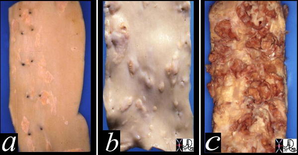
| This image shows three pathological specimens of the aorta. In the first image minimally raised fatty streaks are noted. (a). In image b, the fibrous capsule causes raised fibrofatty nodules, while in c, there gas been rupture of the plaques, with friable atheromatous plaques abound. Courtesy Henri Cuenoid MD 13420c CVS artery aorta atheroscleosis atheroma fatty streaks fibro |
Infiltration of Fat and Fibrous Tissue
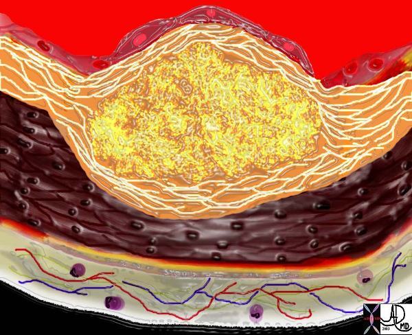
Addition of Fbrous Tissue Creates a Fibrofatty Plaque |
| The diagram shows the atherosclerotic lesion in the subepithelial layer of the intima which is bulging both toward the media and toward the lumen. There is a central core of fat and necrotic debris, surrounded by fibrous elements which give the plaque its hardness to the feel. The accumulation of fibrous tissue heralds an advanced atherosclerotic lesion. 33801b
Courtesy Ashley Davidoff MD. code heart artery atherosclerosis Davidoff art |
|
Irregular Walls with Focal Areas of Thickening |
| 48363 descending thoracic aorta fx aortic ulcer fx atherosclerosis atheroma fx penetrating ulcer CTscan Courtesy Ashley Davidoff MD |

Laying Down of Dystrophic Calcification
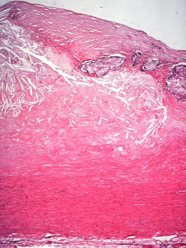
Atherosclerosis of the Aortic Wall |
| 13316 13314 13313 aorta aortic wall elastic layer intima plaque complex lesion atherosclerosis atheroma complex lesion histopathlogy Courtesy Dr Isabelle Joris |
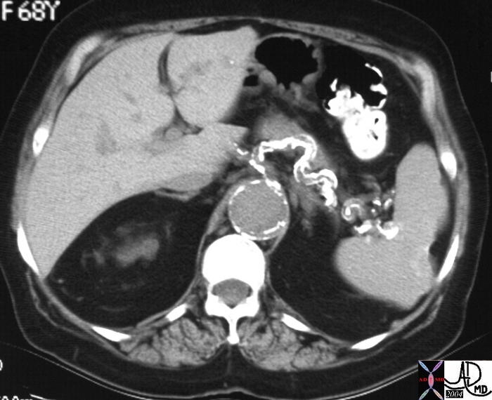
Atherosclerotic Calcification of the Splenic Artery and Aneurysmal Aorta |
| 22014b spleen + fx irregular scar wedge defect calcification + dx chronic splenic infarction + imaging radiology CTscan splenic artery calcified calcification serpigenous atherosclerosis abdomen aorta AAA |