
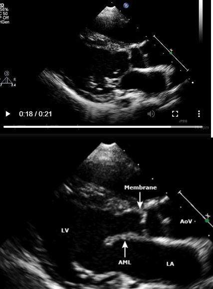
appear to arise from the membranous septum, although they may arise from a thickened muscular ridge slightly further down into the ventricle
Attachments to the anterior leaflet of the mitral valve are frequently seen.
- rare in newborns and infants.
- progresses over time s
- ?acquired
- ? due to thickening and scarring from turbulence caused by an underlying LVOT abnormal architecture.
- ?acquired
Courtesy Up to Date

LV angiogram in RAO projection shows a thin subaortic membrane (arrows)
Ashley Davidoff
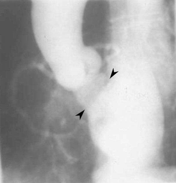
LV angiogram in LAO projection shows a thin subaortic membrane (arrows)
Ashley Davidoff

White blood imaging in the coronal plane shows a subaortic membrane in the LV outflow tract
Ashley Davidoff
86548
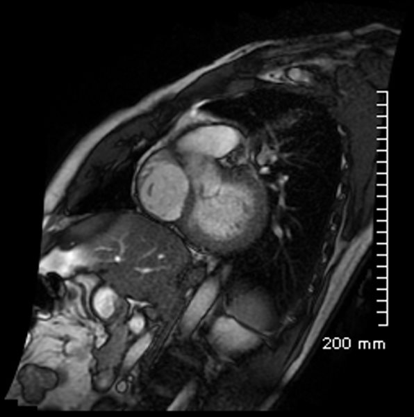
White blood imaging in the sagittal plane shows a subaortic membrane in the LV outflow tract
Ashley Davidoff
86551
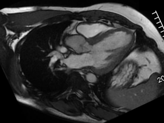
White blood imaging in the axial plane shows a subaortic membrane in the LV outflow tract
Ashley Davidoff
86553