Historical Perspectives
The Common Vein Copyright 2007
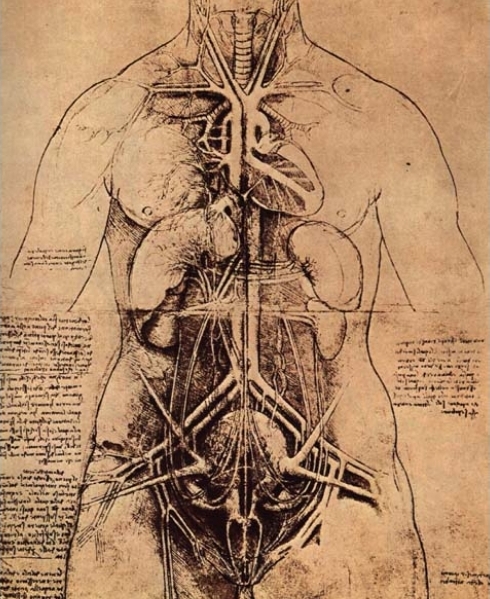
Hippocrates, the Father of Medicine, wrote, “The vessels which spread themselves over the whole body, filling it with spirit, juice, and motion,are all of them but branches of an original vessel. I protest, I do not know where it begins or where it ends, for in a circle there is neither a beginning nor an end.” Although Hippocrates didn’t have the opportunity to view the body with today’s imaging modalities, the assumptions that he made in 470 B.C. were quite accurate. In the course, we analyze one of the major structures of this mysterious circle – the aorta.
Diocles of Carystus (400 BC) is credited with distinguishing the aorta from the cava, but it is probable that this was previously recognized. Aristotle (384-322BC) is given credit for coining the term “aorta” when he distinguished it from the right sided vena cava in animals, but he confused the vena cava and the pulmonary artery. The word aorta is derived from the Greek word aeirein which means to lift.
Erasistratus (300 BC) further theorized that there was a dual circulation from the liver to the heart (by arteries) and from the heart to the lungs. (by veins). He believed that the aorta and arteries transported air from the lungs to the rest of the body.
Galen (130 AD)proposed that blood moved out of the heart via arteries while simultaneously leaving the liver via veins.
In 1557, Vesalius differentiated aneurysms of the thoracic from those of the abdominal aorta.
William Harvey made an essential conceptual advance by describing the circulation in his classic – De Motu Cordis in 1628.
In 1761, “aortitis” was described by Morgagni.
Atherosclerotic disease of the aorta has been demonstrated in Egyptian mummies studied in the 19th century and has continued to be investigated with the evolving technologies.
In 1898 A.D., archaeologists discovered the mummified body reportedly of Mernephtah, (1224-1214 B.C.)in the “Valley of Kings” nea Cairo. Mernephtah was apparently the Pharaoh who chased Moses and the people of Israel through the Red Sea. His calcified aorta with bone like plaques was shown at a meeting of the Royal Society of Medicine in 1909.
In 1819 Laennec invented the stethoscope.
In 1826, the same Laennec first described “dissecting” aortic aneurysms.
In 1868, Allbutt described syphilitic arteritis.
In 1911, arteriosclerosis was found by Sir Armand Ruffer in the aortas of 3000 year old Egyptian mummies.
In 1929 radiologic demonstration of the aorta was seen after injection of radiopaque material by
Lamas Pereira-Caldas de los Santos.
In 1938, Ickikawa was the first to perform abdominal aortography in man.