Takayasu’s arteritis is an inflammatory disease of the aorta and its first order branches
It is caused by intimal proliferation and fibrosis along with fibrous scarring and degeneration of the elastic fibers of the media of the aorta and large arteries. The M:F is 1:8. Typical age of onset is in teenage years.
As a result of the inflammatory process, –
vaso vasorum are destroyed
necrotizing and obliterative segmental,
panarteritis
the adventitia becomes thickened and the
large-vessel
late complications
Localized aneurysm formation,
post-stenotic dilatation and
calcification in the arterial walls are . The process most often involves the arch and its major branches
The clinical diagnosis is suspected when a teenage patient presents with loss of pulses or ischemic parestesias. Non specific findings include fever, malaise, night sweats, arthralgias, fatigue and occasionally pain and tenderness over the affected arteries. Imaging is best accomplished with CTA or MRA and for the smaller vessels angiography is still a useful modality.
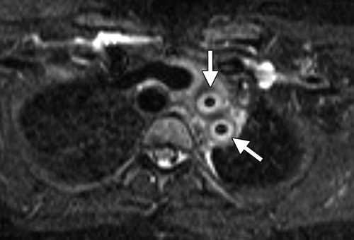
“Aortic wall thickening, which has been described as a “double ring” appearance at contrast-enhanced CT, is the typical finding in the early stage, with a poorly enhanced internal ring (the swollen intima) and an enhancing outer ring (the inflamed media and adventitia) (9).” (Restrepo)
Treatment options include glucocorticoids may relieve systemic symptoms and surgical treatments may be needed for late complications Morbidity and mortality depend on the presence or absence of severe complications such as retinopathy, aortic regurgitation, secondary hypertension or aortic aneurysms There is 97% survival over 7 years in uncomplicated disease and 59% in patients with complications
Smooth Narrowing – Takayasu’s Aortitis
20354b01 14 year old male artery thoracic aorta fx smooth narrowing of isthmus of aorta Takayasu’s aortitis angiography angiogram Courtesy Ashley Davidoff MD
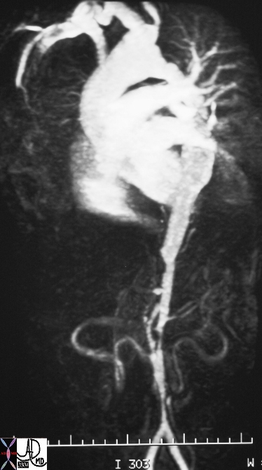
Multicentric and Diffuse Irregular Narrowing of the Istmus and Abdominal Aorta
Takayasu’s Arteritis
In this patient, the MRI shows narrowing of the abdominal aorta segmentally and diffusely in this patient who has Takayasu’s arteritis and aortitis – an inflammatory condition affecting the aorta and large arteries. 16917b Courtesy Ashley Davidoff MD
Takayasu’s Arterirtis
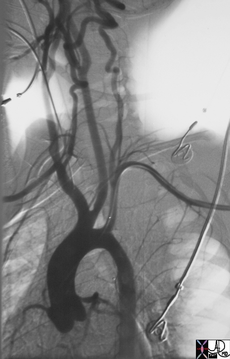
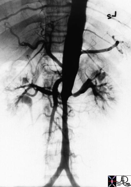
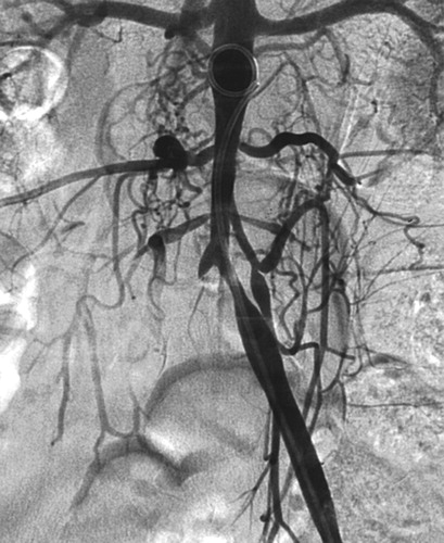
The series of images are from the angiogram of a 14 year old female who presented with seizures and an elevated blood pressure. Images a and b show multiple stenoses within the carotids best seen at the level of the bifurcation into external and internal arteries. In addition in b, the aortic arch shows non critical narrowing just after the origin of the left common carotid vessel. Note that the right subclavian artery is not seen and presumably is accluded at its origin. The abdominal angiogram shows a significant narrowing of the left renal artery with post stenotic dilitation, and stenotic disease in the infrarenal abdominal aorta. The multicentric nature of the disease in a young female is athognomonic of Takayasu’s arteritis. 35155c Courtesy of Laura Feldman MD. code CVS artery aorta arteritis inflammation Takayasu’s carotid thorax arch renal abdomen pulseless
—
Takayasu arteritis in a 13-year-old girl. (a) Image from conventional angiography shows concentric infrarenal narrowing of the aorta and left common iliac artery and occlusion of the proximal right common iliac artery. Radiographics Restrepo 2011
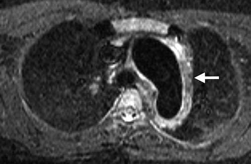
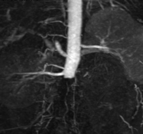
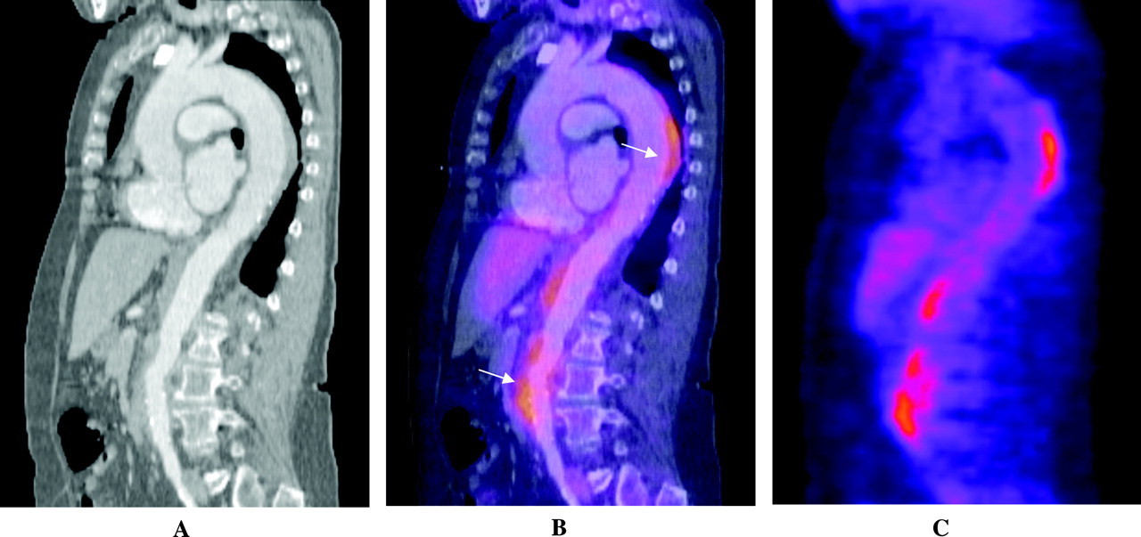
Takayasu arteritis in a 22-year-old woman. Contrast material–enhanced CT image shows extensive thickening of the aortic wall that involves the entire thoracic aorta (arrows). There is an area of ulceration in the anteromedial wall of the descending aorta (arrowhead). Radiographics Restrepo 2011Takayasu arteritis in a 20-year-old woman. Short inversion time inversion-recovery MR images show abnormal arterial wall thickening and edema involving the arch vessels (arrows in a) Radiographics Restrepo 2011Findings similar to the double ring appearance seen at CT. The wall thickening and edema are better seen in the left carotid and left subclavian arteries. Radiographics Restrepo 2011Aortic interruption in a 27-year-old man with Takayasu arteritis. Image from MR angiography shows complete occlusion of the mid abdominal aorta, occlusion of the left renal artery, and multiple retroperitoneal collateral vessels. Radiographics Restrepo 2011Combination of 18F-FDG PET and CTA for assessment of Takayasu arteritis. Shown are sagittal plane contrast-enhanced CTA images of the thoracic and abdominal aorta (A), 18F-FDG PET-CTA overlay (B), and 18F-FDG PET alone (C). Areas of FDG uptake, consistent with inflammation, can also be found, visualized in the descending thoracic and abdominal aorta. Image courtesy of Dr Paul Schoenhagen, Departments of Radiology and Cardiovascular Medicine, Cleveland Clinic Foundation Circulation 2008 Gornick and Creager
References
“Takayasu Arteritis.” Medline Plus. National Library of Medicine, n.d. Web. 14 June 2012. <http://www.nlm.nih.gov/medlineplus/ency/article/001250.htm>.