
Abdominal Aorta
The Common Vein Copyright 2007
Ashley Davidoff MD
Jessica Humphries
Definition
The abdominal aorta is part of the circulatory system and is the final segment of the aorta.
It begins at the end of the thoracic aorta, terminating by bifurcation into the common iliac arteries, and is structurally characterized by its tubular structure, elasticity, and gradually tapering diameter.
The function of the abdominal aorta is to transport blood at a given mean arterial pressure to the systemic arteries, including the mesenteric, splenic, hepatic, pancreatic, renal, gonadal, and iliac vessels.
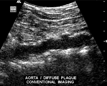
Common diseases of the abdominal aorta include atherosclerosis and aneurismal disease.
Commonly used diagnostic procedures for atherosclerosis include ultrasounds, CT scans, and MRI scans while only a clinical exam is necessary in diagnosing aneurismal disease.
These diseases are usually treated with minimally invasive surgical techniques, as well as open surgery.
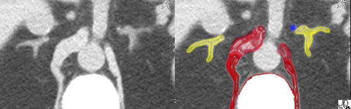 Aortic Hiatus Aortic Hiatus |
| The crura of the diaphragm (maroon overlay) can be seen as muscle bundles alongside the vertebral body and surrounding the aorta. The limbs of the adrenal gland (yellow overlay)should not be thicker than the crura. The aortic hiatus is created by the two crura and marks the end of the thoracic aorta and the beginning of the abdominal aorta
(Image courtesy of Ashley Davidoff M.D.) adrenal gland bilateral nodules adenomas neoplasm benign CTscan imaging radiology 39502 |
The abdominal segment originates at the aortic hiatus of the diaphragm at the plane of the thoracic and lumbar vertebral junction. The aorta is surrounded by the right and left crura at this stage. The abdominal aorta is structurally characterized as a tapering tube, composed of muscular and elastic tissue. Along its course, the abdominal aorta gives rise to several large branches including the celiac axis, the superior mesenteric artery (SMA), and the inferior mesenteric artery (IMA arising 3 to 4 cm above the bifurcation. The renal arteries arise from the lateral border of the aorta between the SMA and IMA branches. The gonadal arteries (testicular and ovarian) are small branches that also arise from the lateral border of the abdominal aorta below the origin of the renals and above the IMA. Common diseases include abdominal aortic aneurysm, (AAA – “triple A”) and atherosclerosis. Diagnostic studies include CT imaging, MRI, aortography and ultrasound Treatment is usually surgical, and with percutaneous techniques such as endoluminal graft insertion becoming more frequently utilized.
It is the artery from which all arteries supplying blood to tissues below the diaphragm are derived.
Parts
Suprarenal Abdominal Aorta
The suprarenal aorta as its name implies is that portion of the abdominal aorta that lies above the renal arteries. Its first branch is the celiac axis which arises anteriorly and which itself gives rise to the arteries of the foregut. The superior mesenteric artery arises about 1 cm distally, supplying structures of the midgut. The suprarenal aorta also supplies a number of smaller arteries, including the inferior phrenic arteries, the suprarenal vessels, and the paired gonadal arteries. Atherosclerosis is common in this segment of the aorta but aneurysmal disease is more common in the ingrarenal segment. When aneurysms occur at the level of the renal arteries or above they present a significant challenge for the surgeon.
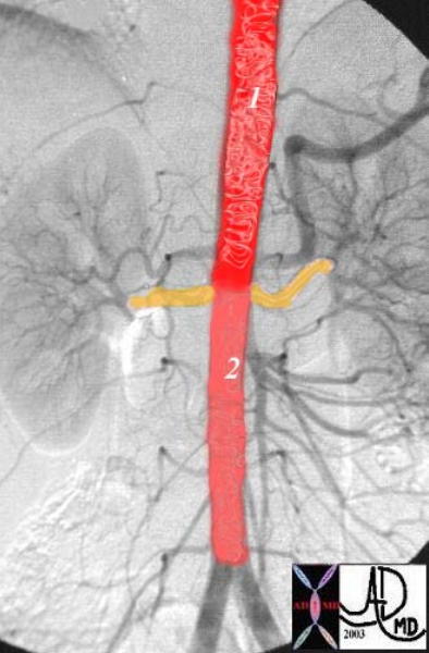
|
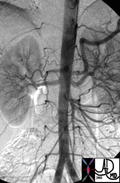
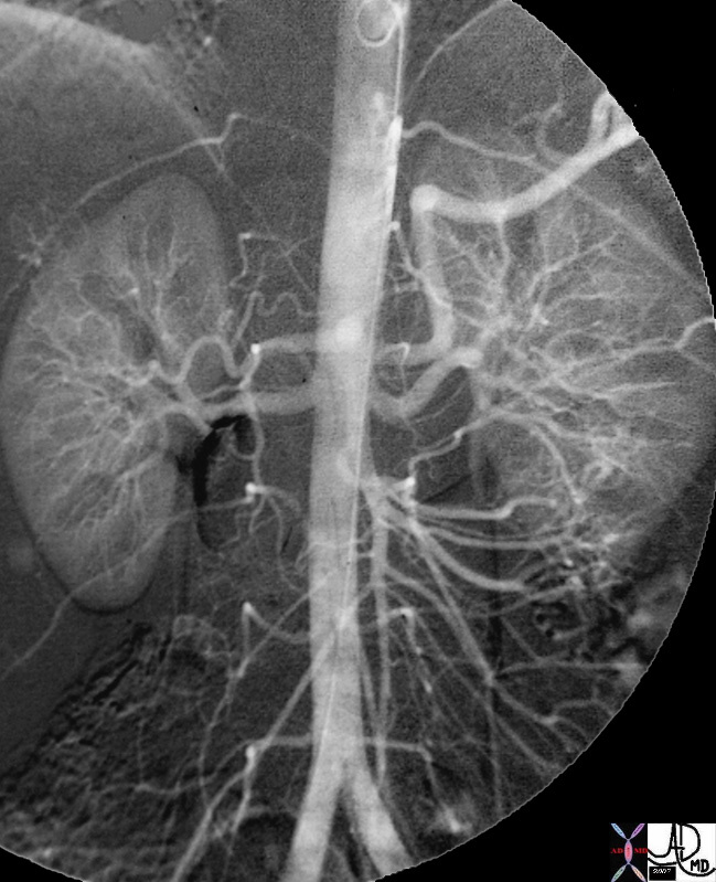
| This overlay of a normal abdominal angiogram shows the two components of the abdominal aorta. The suprarenal component, (1) is slightly larger than the infrarenal component (2). The renal arteries, (in orange overlay,=) take 20% of the cardiac output and hence a large portion of the aortic blood flow is “unloaded” at this level.
Courtesy Ashley Davidoff MD. 24877d code CVS aorta abdomen normal suprarenal infrarenal |
 Suprarenal and Infrarenal Abdominal Aorta
The infrarenal aorta as its name implies lies below the origin of the renal arteries. It is located between the top of L2 and the bottom of L4. It gives rise on its anterior surface to the inferior mesenteric artery, supplying structures of the hindgut, and from both posterolateral walls to a number of lumbar vessels, and to a middle sacral artery at the level of the aortic bifurcation. Common diseases include abdominal aortic aneurysm, (AAA – “triple A”) and atherosclerosis. Diagnostic studies include CT imaging, MRI, aortography and ultrasound Treatment is usually surgical, with percutaneous techniques such as endoluminal graft insertion becoming more frequently utilized.
Size
The abdominal aorta gives off many branches and diminishes rapidly in size from about 2.5cms to about 1.75 cm. in diameter, finally terminating into the two common iliac arteries. In females it is slightly smaler Cristina see if you can get more exact numbers
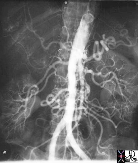
Caliber Change of the Abdominal Aorta Well Demonstrated |
| 25661.800 aorta abdomen abdominal normal anatomy size celiac axis renal arteries Angiogram angiogaphy Courtesy Ashley DAvidoff MD |
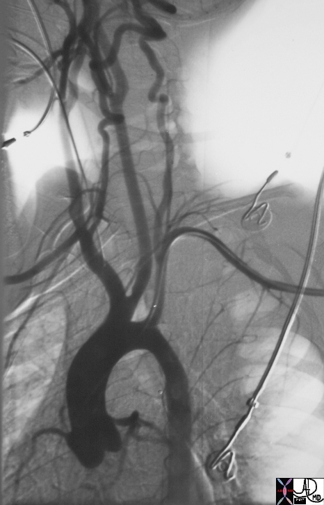
Shape
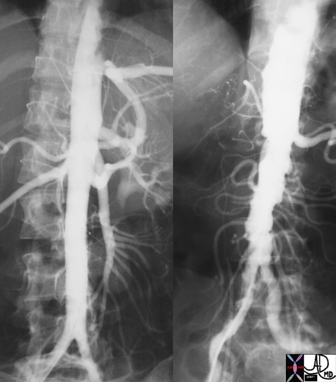
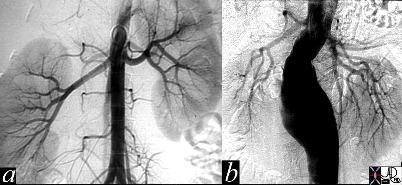
The abdominal aorta is a straight tube in th young and becomes elongated tortuous and enlarges with age
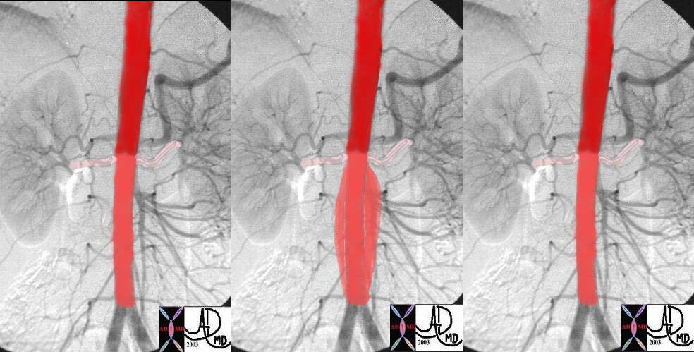
The abdominal aorta is a straight tube in th young and becomes elongated tortuous and enlarges with age |
| These digitally subtracted angiograms show progression from the straight tube in the first image becoming mildly atherosclerotic and tortuous in the middle image, and finally becoming a fusiform infrarenal abdominal aortic aneurysm. There is mild ectasia of the right common iliac artery. Courtesy Ashley Davidoff MD 24877b26968 24590 code aorta abdomen aneurysm angiogram AAA |
The abdominal segment originates at the aortic hiatus of the diaphragm at the plane of the thoracic and lumbar vertebral junction. The aorta is surrounded by the right and left crura at this stage. The abdominal aorta is structurally characterized as a tapering tube, composed of muscular and elastic tissue. Along its course, the abdominal aorta gives rise to several large branches including the celiac axis, the superior mesenteric artery (SMA), and the inferior mesenteric artery (IMA arising 3 to 4 cm above the bifurcation. The renal arteries arise from the lateral border of the aorta between the SMA and IMA branches. The gonadal arteries (testicular and ovarian) are small branches that also arise from the lateral border of the abdominal aorta below the origin of the renals and above the IMA. Common diseases include abdominal aortic aneurysm, (AAA – “triple A”) and atherosclerosis. Diagnostic studies include CT imaging, MRI, aortography and ultrasound Treatment is usually surgical, and with percutaneous techniques such as endoluminal graft insertion becoming more frequently utilized.
It is the artery from which all arteries supplying blood to tissues below the diaphragm are derived.
Parts
Suprarenal Abdominal Aorta
The suprarenal aorta as its name implies is that portion of the abdominal aorta that lies above the renal arteries. Its first branch is the celiac axis which arises anteriorly and which itself gives rise to the arteries of the foregut. The superior mesenteric artery arises about 1 cm distally, supplying structures of the midgut. The suprarenal aorta also supplies a number of smaller arteries, including the inferior phrenic arteries, the suprarenal vessels, and the paired gonadal arteries. Atherosclerosis is common in this segment of the aorta but aneurysmal disease is more common in the ingrarenal segment. When aneurysms occur at the level of the renal arteries or above they present a significant challenge for the surgeon.
| Suprarenal and Infrarenal Abdominal Aorta |
| This overlay of a normal abdominal angiogram shows the two components of the abdominal aorta. The suprarenal component, (1) is slightly larger than the infrarenal component (2). The renal arteries, (in orange overlay,=) take 20% of the cardiac output and hence a large portion of the aortic blood flow is “unloaded” at this level.
Courtesy Ashley Davidoff MD. 24877d code CVS aorta abdomen normal suprarenal infrarenal |
Infrarenal Abdominal Aorta
The infrarenal aorta as its name implies lies below the origin of the renal arteries. It is located between the top of L2 and the bottom of L4. It gives rise on its anterior surface to the inferior mesenteric artery, supplying structures of the hindgut, and from both posterolateral walls to a number of lumbar vessels, and to a middle sacral artery at the level of the aortic bifurcation. Common diseases include abdominal aortic aneurysm, (AAA – “triple A”) and atherosclerosis. Diagnostic studies include CT imaging, MRI, aortography and ultrasound Treatment is usually surgical, with percutaneous techniques such as endoluminal graft insertion becoming more frequently utilized.
Size
The abdominal aorta gives off many branches and diminishes rapidly in size from about 2.5cms to about 1.75 cm. in diameter, finally terminating into the two common iliac arteries. In females it is slightly smaler Cristina see if you can get more exact numbers
| Caliber Change of the Abdominal Aorta Well Demonstrated |
| 25661.800 aorta abdomen abdominal normal anatomy size celiac axis renal arteries Angiogram angiogaphy Courtesy Ashley DAvidoff MD |
Shape
The abdominal aorta is a straight tube in th young and becomes elongated tortuous and enlarges with age
| The abdominal aorta is a straight tube in th young and becomes elongated tortuous and enlarges with age |
| These digitally subtracted angiograms show progression from the straight tube in the first image becoming mildly atherosclerotic and tortuous in the middle image, and finally becoming a fusiform infrarenal abdominal aortic aneurysm. There is mild ectasia of the right common iliac artery. Courtesy Ashley Davidoff MD 24877b26968 24590 code aorta abdomen aneurysm angiogram AAA |
The abdominal aorta is a staright tube in th young and becomes elongated tortuous and enlarges with age
Position and Relations
The abdominal aorta begins at the diaphragm in front of the last thoracic vertebra and descends in front of the vertebral column to the fourth lumbar vertebra, slightly to the left of the middle line.
 Normal Abdominal Aorta in the Central Retroperitoneum Normal Abdominal Aorta in the Central Retroperitoneum |
| 60763c01 abdomen abominal aorta relations left renal vein renal artery arteries anterior pararenal space central retroperitoneum lumbar spine CTscan Courtesy Ashley Davidof mD |
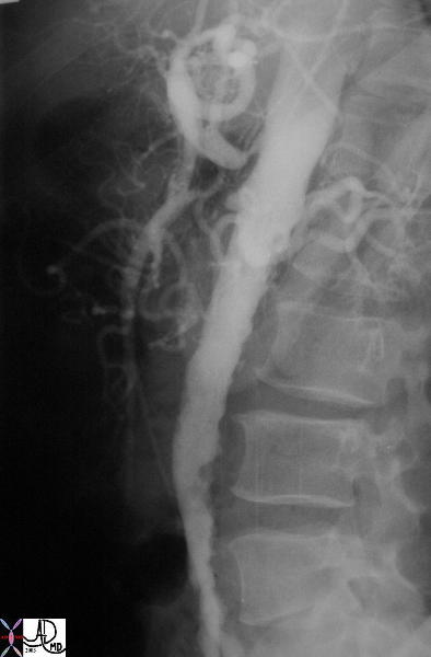 The Aorta Hugs the Anterior Border of the Lumbar Spine The Aorta Hugs the Anterior Border of the Lumbar Spine |
| The lateral projection of the digital angiogram shows mild to moderate infrarenal atherosclerosis characterised by luminal irregularity. There is also mild lumenal narrowing of the affected infrarenal segment. The suprarenal aorta is smooth and within normal limits. The celiac axis has a high grade sternosis and the SMA shows a subtotal occlusion. (See also 35059 35060) 35061 Courtesy Laura Feldman MD. code abdomen aorta artery atherosclerosis celiac axis occlusion SMA stenosis. |
Character
Elastic nature
 Elastic Nature Elastic Nature |
| 24877c This angiogram shows a theoretical changes of the aortic wall during diastole (a) sytolic expansion (b) and return to normal in diastole (c) In systole (b) the suprarenal artery is expanded by the pulse but is relatively decompressed by the the low resistance and high flow renal arteries. The infrarenal aorta is relatively more expanded in systole (b) since the iliac arteries offer a relative resistance. This increased resistance causes the elastic tissue in the aorta to stretch (b) so that the recoil in diastole (c) results in a sustained forward moving force assisting the blood to get to their most distal destination – the feet. When the aorta starts to lose its elasticiy the recoil of systole gradually is weakened so that the aorta does not return to its normal diameter after each systolic expansion. Over many years this lack of recoil is progressive so that the resulting wall is weaker and dilated, until an aneurysm is formed. A small infrarenal fusiform aneurysm is seen in images d(diastole), e, (systole) and f, diastole. Some recoil is still present, as seen by a systolic dilatation (e) with slight decrease in diameter during diastole (f) Courtesy Ashley Davidoff code CVS artery aorta AAA infrarenal circulatory elasticity recoil |
Applied Biology
Atherosclerosis
Degenerative Inflamma diis Cause Result dx Rx
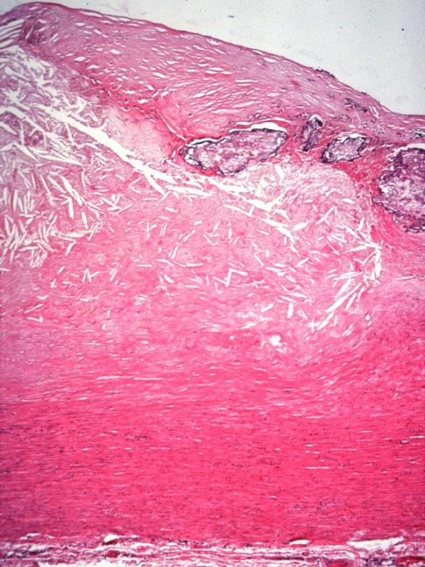
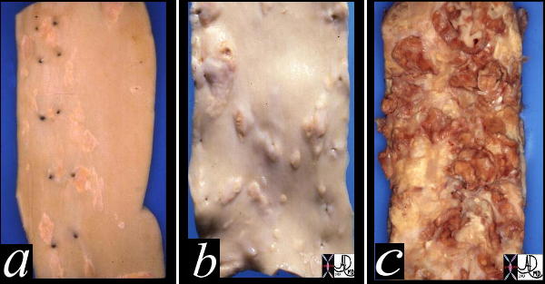 Fatty Streaking, Raised Fibrofatty plaques, and Porridge like Friable Atherpmatous Plaque Fatty Streaking, Raised Fibrofatty plaques, and Porridge like Friable Atherpmatous Plaque |
| This image shows three pathological specimens of the aorta. In the first image minimally raised fatty streaks are noted. (a). In image b, the fibrous capsule causes raised fibrofatty nodules, while in c, there gas been rupture of the plaques, with friable atheromatous plaques abound.
Courtesy Henri Cuenoud MD 13420c CVS artery aorta atheroscleosis atheroma fatty streaks fibro-fatty plaque |
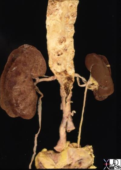 Atherosclerosis and Left Sided Renal Artery Stenosis Atherosclerosis and Left Sided Renal Artery Stenosis |
| 13417 aorta abdomen iliac artery fx atherosclerosis kidney fx small dx RAS renal artery stenosis grosspathology Courtesy Ashley Davidoff MD |
Normal Aorta and Atherosclerosis |
| 13417 aorta abdomen iliac artery fx atherosclerosis Courtesy Philips Medical Systems AO1A.JPG AO2A.JPG |
Atherosclerosis of the Aortic Wall |
| 13316 13314 13313 aorta aortic wall elastic layer intima plaque complex lesion atherosclerosis atheroma complex lesion histopathlogy Courtesy Dr Isabelle Joris |
Normal and Severe Atherosclerosis |
| 18485c01 aorta artery hepatic artery renal artery splenic artery superior mesenteric artery SMA kidney severe atheroscleroris atheroma occluded renal artery spleen liver normal anatomy angiogram angiography Davidoff MD |
Abdominal Aortic Aneurym
Degenerative Disease related to atheroscleorosis Cause loss of elasticity Result Dx and Rx
|
Abdominal Aneurysm |
| Courtesy Philips Medical Systems 1153BH~1.JPG |
Normal and AAA |
| 11976c01 aorta abdomen abdominal aorta renal arteries kidney fx normal AAA abdominal aortic aneurysm horseshoe kidney angogram angiography lumbar arteries Davidoff MD |
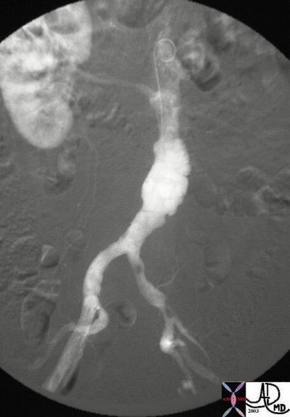
AAA with horseshoe kidney |
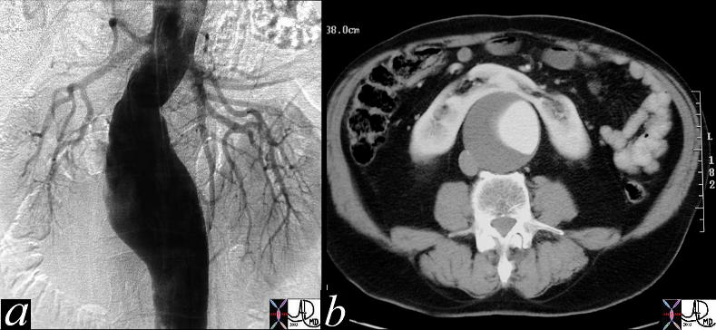
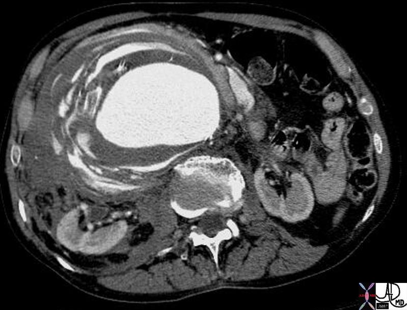
| This digital angiogram of an infrarenal, fusiform abdominal aortic aneurysm, shows an expanded and tortuous aortic lumen. The contrast column represents the luminal diameter and not the true size of the aneurysm. The size is best evaluated on cross sectional imaging where the outside edges of the wall are seen to best advantage. (b). Note there is a large thrombotic component only appreciated on the CT scan. A horseshoe kidney is vaguely outlined on the angiogram, but is quite obvious on the CT scan. This is an essential piece of information for the surgeon, and makes the surgery that much more difficult.
Courtesy Ashley Davidoff 11976c code CVS aorta abdomen AAA kidney horseshoe |
|
AAA with horseshoe kidney |
| This digital angiogram of an infrarenal, fusiform abdominal aortic aneurysm, shows an expanded and tortuous aortic lumen. The contrast column represents the luminal diameter and not the true size of the aneurysm. The size is best evaluated on cross sectional imaging where the outside edges of the wall are seen to best advantage. (b). Note there is a large thrombotic component only appreciated on the CT scan. A horseshoe kidney is vaguely outlined on the angiogram, but is quite obvious on the CT scan. This is an essential piece of information for the surgeon, and makes the surgery that much more difficult.
Courtesy Ashley Davidoff 11976c code CVS aorta abdomen AAA kidney horseshoe |
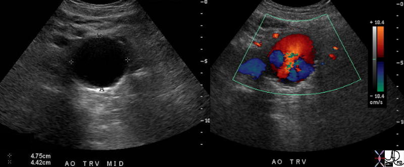
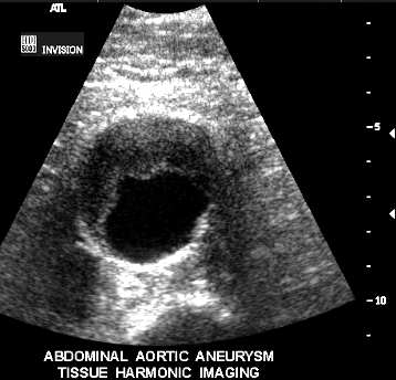
Turbulence in an Abdominal Aortic Aneurysm |
| 37669c01 aorta abdomen abdominal aort AAA enlarged aneurysm abdominal aortic aneurysm turbulent flow USscan color flow doppler Courtesy Ashley Davidoff MD |
Smooth Narrowing of the Lumen Due to Aneurysmal Disease |
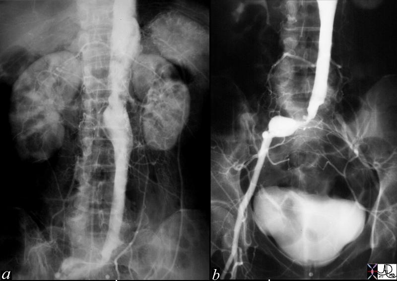
| This angiogram of the abdominal aorta (a) and iliac arteries (b), shows an unusually straight and narrowed infrarenal aorta indicative of thrombus in the wall of an abdominal aortic aneurysm. In addition there is an aneurysm of the right common iliac artery and a subtotal occlusion of the left common iliac artery. Note the left kidney is small and there is a wedge shaped defect in the upper and lateral aspect of the kidney indicative of an infarct, probably embolic in origin. Courtesy Laura Feldman MD. 36005c code abdominal aorta aneurysm artery iliac stenosis occlusion kidney infarct wedge embolus |
Aortic Rupture of AAA
Definition Cause Complication of AAA usially when > 5.5 cms Result catasstrophic Dx CT RX OR emergency
Ruptured AAA |
| 18269 aorta abdomen AAA aortic aneurysm abdominal aorta retroperitoneum fx retroperitoneal hematoma fx active hemorrhage fx perinephric hematoma anterior pararenal space perirenal space posterior pararenal space hemorrhage dx rupture abdominal aortic aneurysm CTscan Davidoff MD fx ruptured AAA |
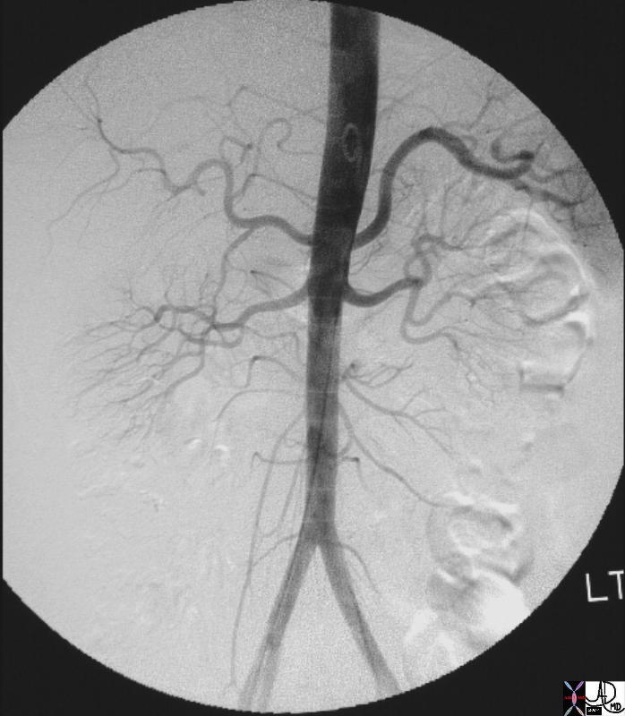
Atherisclerotic Occlusion of the Aorta
Definition Cause Result Dx RX
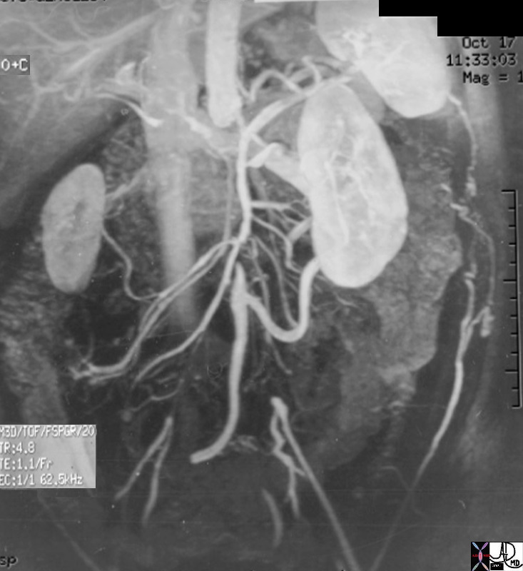
Athrosclerotic Occlusion of the Aorta |
| 14646.803 aorta kidney size hypertrophy atrophy artery superior mesenteric artery arc of Drummond IMA inferior mesenteric artery collaterals renovascular disease occluded aorta internal iliac artery fx occluded occlusion small compensatory hypertrophy collaterals MRA MRIscan Davidoff MD |
Descending Aortic Dissection
Descending Aortic Dissection
Type – mechanical disorder – ause Cause Resuly Dx and Rx
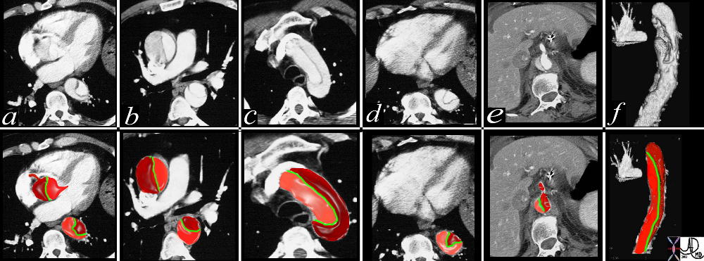
Type C Dissection |
|
This combination of images from a CTscan through the thorax, reveals an aortic dissection starting at the level of the aortic valve and extending into the abdomen with involvement of the celiac axis. The true lumen is overlaid in pink while the false lumen is over;laid in a darker red. The green structure is the intimal flap. Flow in the false lumen is slightly delayed and slower as seen by the lower density of the contrasyt within it. Courtesy Ashley Davidoff MD. 19412c02 code CVS artery AO aorta dissection fx crescent |
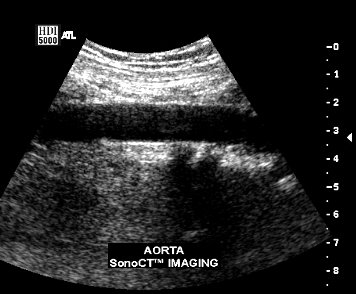
Descending Thoracic Aorta – Dissection |
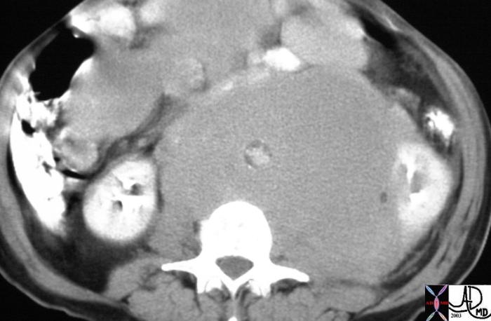
| This cross sectional image of the mid abdmen shows an aorta with an expanded diameter, which in this case is associated with an extremely small lumen. Note the wall of atherosclerotic calcification is on the inside of the soft tissue surrounding it. The case represents non-Hodgkins abdominal lymphoma that masquerades as an abdominal aortic aneurysm. The positioning of the calcification is key to this recognition
. Courtesy Ashley Davidoff MD 15657 code CVS aorta large lyphoma radiologists and detectives |
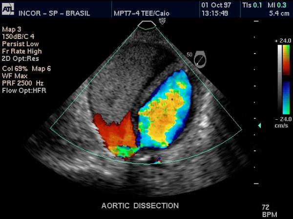
Descending Thoracic Aorta – Dissection |
| Doppler US of the descending thoracic aorta showing flow in the true lumen (color) and no flow in the thrombosed lumen (gray echoes) in this patient with aortic dissection. Courtesy Philips Medical Systems 33166 |
Trauma
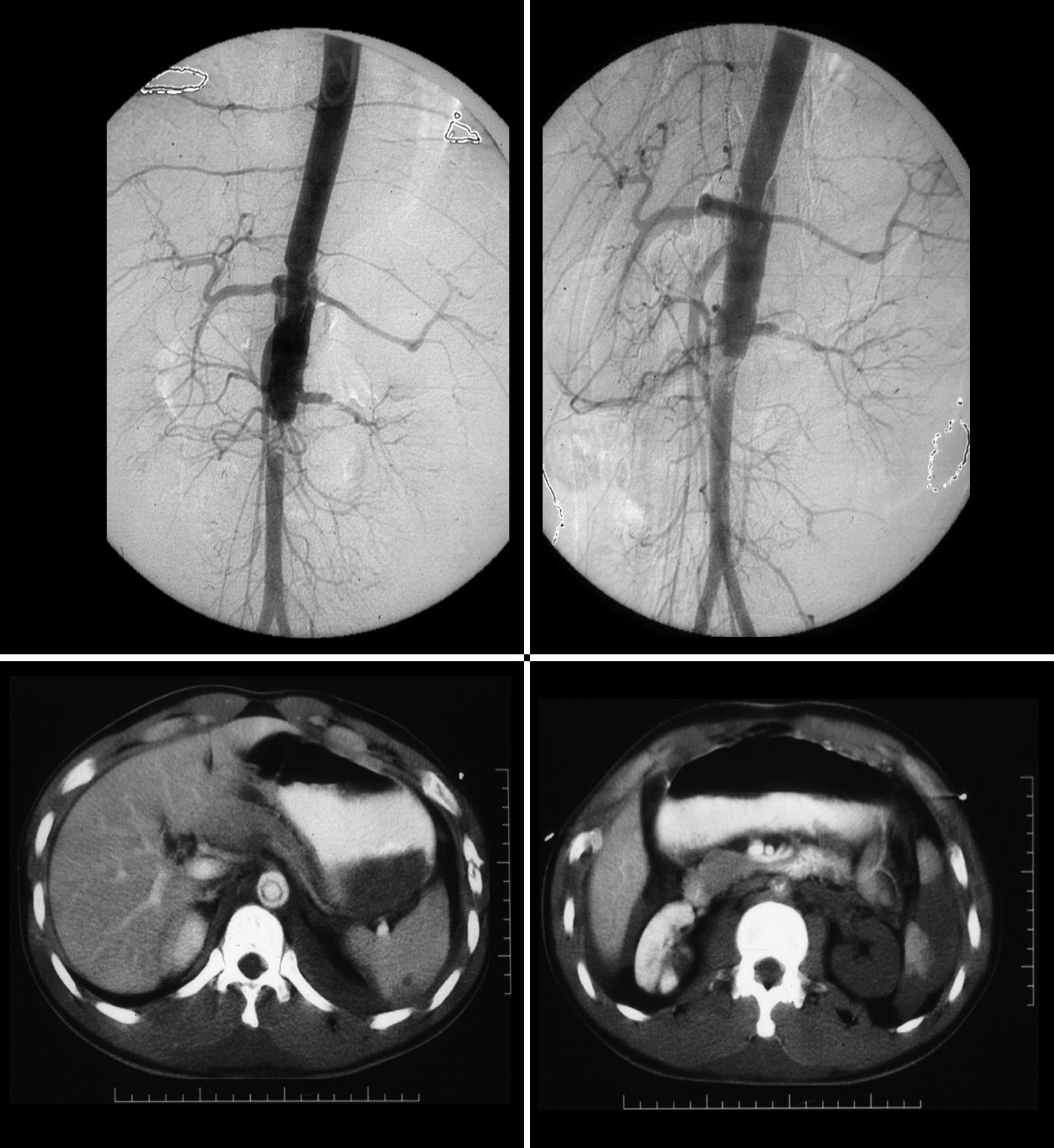
Traumatic Dissection of the Abdominal Aorta wit Loss of Perfusion of the Left Renal Artery |
| These images represent a traumatic dissection of the abdominal aorta caused by a compression injury to the abdomen. The angiograms in a and b shows what appears to be a complex tear of the aorta with a dissection and then a subtotal obstruction of the aorta distally. The CTscan shows (c) the dissection to better effect, with a subtotal narrowing of the aorta distally. Note also the injury of the left renal artery on the angiogram as well as the lack of perfusion of the left kidney on the CT scan. Courtesy Ashley Davidoff MD. 14734c code aorta abdomen trauma dissection |
Takayasus’s Aortitis
An infllammatory diseas Cause Result Dx Rx
Takayasus’ Aortitis Proximal Descending Thoracic Aorta |
| 20354b01 14 year old male artery thoracic aorta fx smooth narrowing of isthmus of aorta Takayasu’s aortitis angiography angiogram Courtesy Ashley Davidoff MD |
Therapy

1680BH~1.JPG