
Congenital Disease
Copyright 2007
Aortic Valve
|
Normal and Thickened Aortic Valve over Time |
| 07953c02 heart cardiac aorta aortic valve fx normal fx thickened fx bicuspid aortic valve fx calcified fx calcification fusion of the intercoronary commisures grossanatomy |
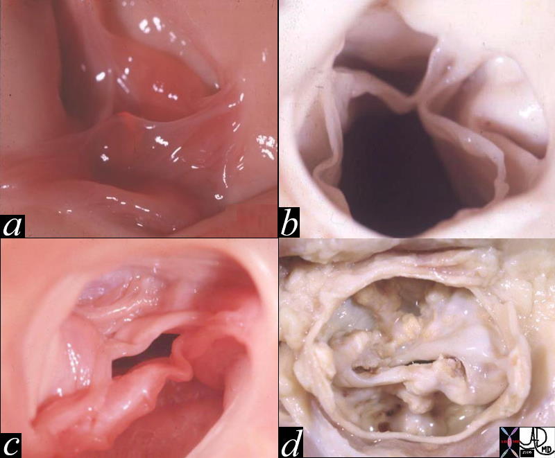
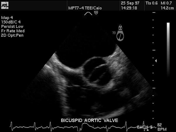
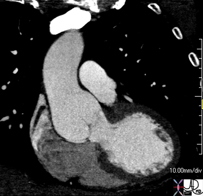
Bicuspid Aortic Valve |
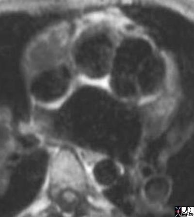
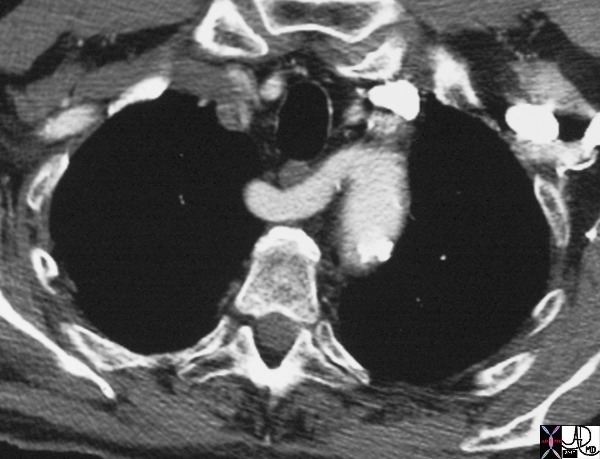
| This gray scale echo of the heart showing a short-axis aorta left atrial view, and demonstrating the aortic valve with two cusps. The patient has a diagnosis of bicuspid aortis valve which is a congenital condition. Courtesy Philips Medical Systems 33169 code cardiac heart echo aorta bicuspid aortic valve congenital imaging cardiac echo |
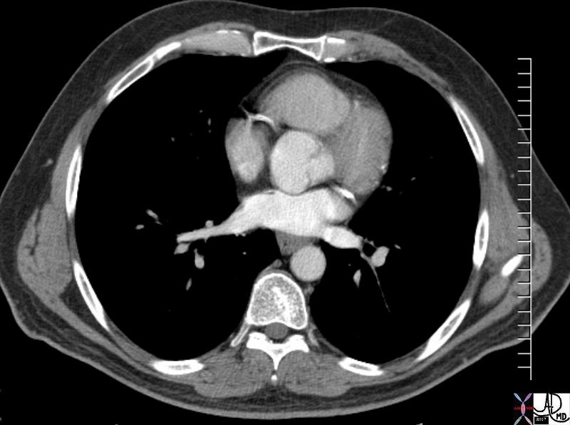
Bicuspid Aortic Valve |
||||
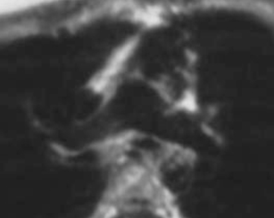
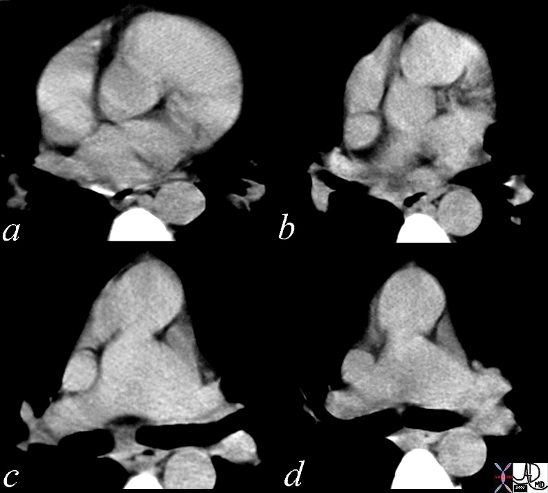
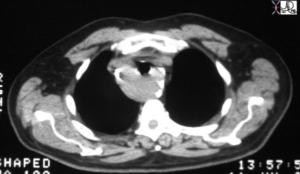
| 72885.800 thorax thoracic aorta aortic valve fx thickened dx bicuspid aortic valve CTscan Courtesy Ashley Davidoff
D Transposition of the Great Vessels
|
Corrected Transposition
 LTGA LTGA |
| 07304b01 anterior aorta posterior pulmonary artery position connection subaortic conus L TGA L TGV L transposition of the great vessels L transposition of the great arteries levo leftward MRI Davidoff MD |
 LTGA LTGA |
| 28999b01 heart cardiac aorta pulmonary artery RVOT conotruncal malformation LTGA L TGV transposition of the great vessels transposition of the geat arteries corrected transposition position connection relation embryology CTscan Davidoff MD 28994 28995 28996 28997 28998 28999 |
|
Right Aortic Arch
|
Supravalvar Aortic Stenosis |
| Hx 4 year female with cocktail pesonality hearyt cardiac artery aorta supravalvular aortic stenosis supravalvar aortic stenosis Williams syndrome William’s syndrome 08233b01 |
Sinotubular Ectasia – Marfan’s Syndrome |
| 72712 thoracic aorta sinus of Valsalva aortic sinuses sinotubular junction of ascending aorta sinotubular ectasia fx dilated CTscan Courtesy Ashley DAvidoff MD 72709 |
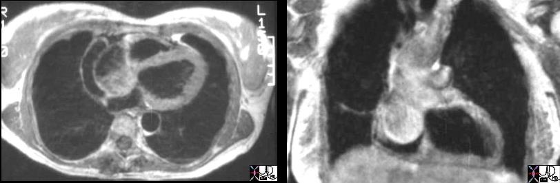
Marfan’s Syndrome Sinus of Valsalva Aneurysm |
| 07974c01 heart cardiac right atrium aortic valve aortic sinus fx right atrial filling defect dx sinus of Valsalva aneurysm of the right aortic sinusprolapsing into the right atrium (RA defect) dx Marfan’s Syndrome MRI T1 weighted Davidoff MD 07973.800 07974c01 07974.800 |
Ascending Aorta
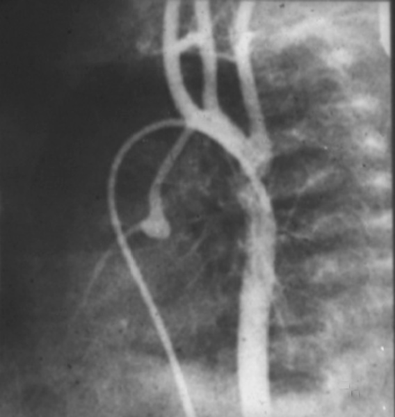
Aortic Valve Atresia in the Setting of Hypoplastic Left Heart Syndrome |
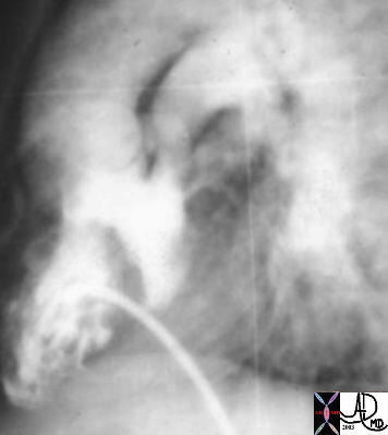
| 00269b02 heart cardiac coronary artery aorta small dx aortic atresia tubular hypoplasia aortic coarctation aortic atresia PDA patent ductus arteriosus angiogram angiogaphy CHD congenital heart disease Davidoff MD 00269b01 00269b02 00269b03 |
Arch
50 year old female with respiratory difficulty trachea bronchi rectus abdominis muscle compression fractures kyphosis dwarf dwarfism right aortic arch tracheomalacia tracheal stenosis rectus abdominis muscle hypertrophy
Courtesy Ashley Davidoff MD TheCommonvein.net
CTscan shape size position character growth 49838c05
Right Aortic Arch |
| 35334 Courtesy of Laura Feldman MD. code aorta arch artery right aortic arch thorax |
Cervical arch
Pseudocoarctation |
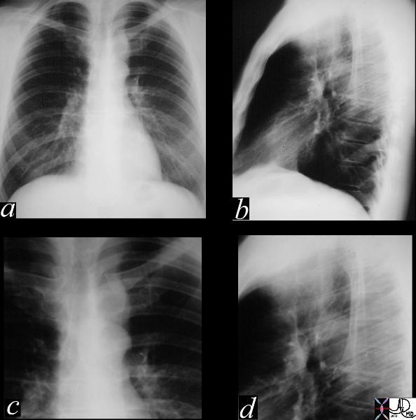
| This series of CXRays P-A (a) lateral (b) and magnified views (c,d) showing an aberrant shape to the aortic knob which is characterised by a “3 sign” reminiscent of a coarctation segment. There is no assocated rib notching nor a pressure gradient and hence the diagnosis is a pseudocoarctation of the thoracic aorta. 35187c01 Courtesy LAura FEldman MD code CVSartery aorta thorax 3 sign peudocoarctation. |
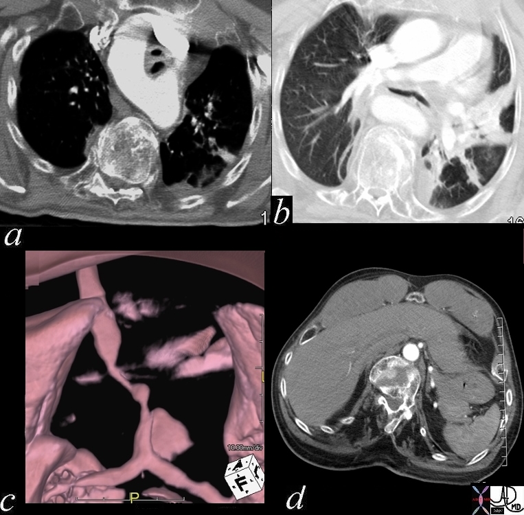
Aberrant Right Subclavian Artery |
| 16376 aorta aberrant origin of right subclavian artery as the last vessel off the left aortic arch congenital growth position esophagus CTscan Davidoff MD |
Isthmus
Patent Ductus Arteriosus |
| 42326.800 aorta pulmonary artery finding PDA patent ductus arteriosus left to right shunt arteriovenous malformation CTscan Courtesy Ashley Davidoff MD |
|
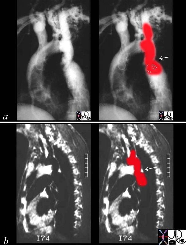
Coartcation of the Aorta |
| This combination of an angiogram and MRI show a focal coarctation of the aorta distal to the dilated left subclavian artery. Note the large internal mammary artery in the angiogram (a) Courtesy Ashley Davidoff MD 00254c02 CVS aorta thorax thoracic aorta coarctation |
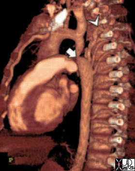
Coarctation CTscan |
| 44146 coarctation of the aorta heart cardiac CTscan MDCT Courtesy Philips Medical |
Aortic Interruption
Congenital – may be in isolation part of the HLHS or in association with other congenital heart diseases dx imaging echo sometimes MRI rx Non invasive techniques and surgical
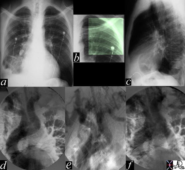
Aortic Interruption at the Level of the Isthmus |
| The most obvious finding in this CXR (a) with pleuro-parenchymal changes is not the most significant. In image (b) the highlighted ribs reveal rib notching characteristic of coarctation of the aorta. The lateral examination (c) in this instance is not helpful. In the early phase of the angiogram(d), there appears to be complete interruption of the aorta with a large left subclavian artery acting as a collateral pathway. The sbsequent images e, and f, show progressive filling of the isthmus and distal thoracic aorta. The coarcatation becomes apparent characterised by a “3” sign. 35107c Courtesy Laura Feldman MD code CVS artery aorta thorax coarctation rib notching bone collateral |