
Shape
Copyright 2007
Introduction
The aorta is a large vascular tube having a nearly round cross section
Thoracic Aorta
keywords aorta aortic valve sinotubular junction ascending aorta aortic arch brachiocephalic artery left common carotid artery left subclavian artery descending aorta normal TCV
Ashley Davidoff MD
47676
Annulus
The annulus is oval
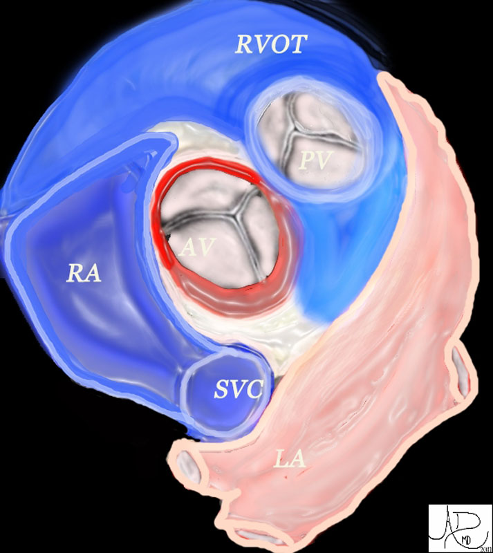
The aortic valve (AV) lies central to many structures including the right ventricular outflow tract (RVOT), pulmonary valve (PV), left atrium (LA), right atrium (RA), and superior vena cava (SVC). aorta from above. In this diagram, the commissures are overlaid in green, the crescent shaped lunulae reflect the free edge of the leaflets and are overlaid in yellow, and the nodules of Arantii are overlaid in orange. The sinuses of Valsalva are like cups and are positioned between the free edges. (gray)
Davidoff art Ashley Davidoff MD TheCommonVein.net 47681Sinuses
The sinuses as their name implies are bulbous.
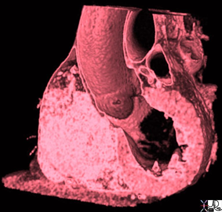
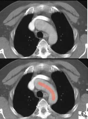
Aortic Bulb, Sinotubular Junction, and Tubular Components of the Ascending Aorta |
| 47823 heart cardiac ascending aorta shape aortic tuck mitral valve to aortic fibrous continuity AV MV normal anatomy CTscan Davidoff MD |
Tubular Portion
The ascending aorta is tubular as its name implies, and it curves gently from the sinotubular junction to the right, reaches almost to the lateral edge of the mediastinum and the curves back to join the beginning of the arch.
Ascending Aorta Bulbous Portion and Tubular Portion |
| 35324b02 thoracic aorta ascending normal Courtesy of Laura Feldman MD code aorta arch artery ascending descending normal thorax |
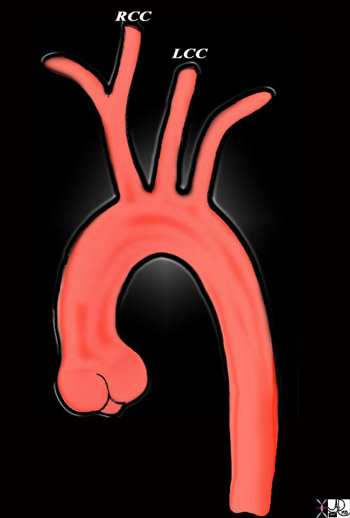
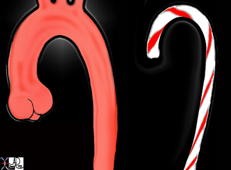
Aortic Arch
The arch has a complex shape with two curves. The first is the more easily understood vertical arch formed as it ascends up and over the left mainstem bronchus. It is often described as being candy cane in shape.
Key Words
candy cane shape aorta aortic valve sinotubular junction ascending aorta aortic arch descending aorta normal
Ashley Davidoff
47677c03
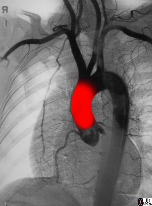
However when viewed in the frontal projection the arch is not quite perfect.
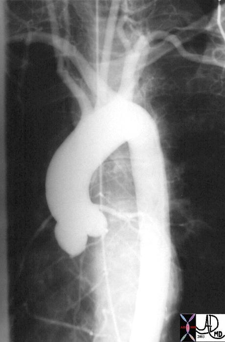
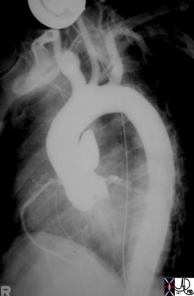
Angle of the Arch in this Projection has an Acute Component |
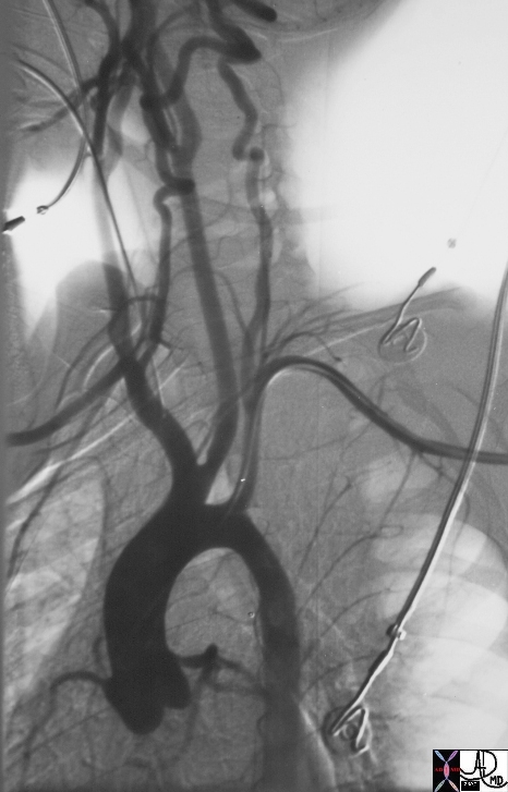
| This angiogram of the normal thoracic aorta is taken in the LAO projection. The thoracic aorta consists of a bulbous portion called the aortic sinus that is the most proximal portion, and is connected to the tubular portion that connects the sinus portion to the aortic arch. The isthmus connects the aortic arch to the descending thoracic aorta.
(see 27185g for overlay image) Courtesy Ashley Davidoff MD 27185 code CVS aorta thorax ascending sinus sinotubular junction tubular AOV arch isthmus descending |
Note that the angle between the arch and descending aorta is too acute.
This aberrancy is due to the a second bed or arch in the aorta as it crosses the the trachea. The arch of the aorta thus forms two curves, one with convexity upward and over the trachea and the second an almost horizontal bend around the trachea first first to the left and then posterior.
Images courtesy of: Ashley Davidoff, M.D. TheCommonVein.net
aorta Philips 026
Thus when you view the arch of a conventional thoracic aortogram, you will be able to understand why it does not look like a perfect vertical arch – it is also bent slightly folded around the trachea and esophagus. In the elderly the aortic knob becomes prominent because of unfolding of the aorta. This means that the second bend flattens making the knob more prominent.

This angiogram of the normal thoracic aorta is taken in the left anterior oblique (LAO) projection. In this projection the shape is not quite a candy cane suggesting some complexity to the arch and known when seen as a three-dimensional structure. Image courtesy of: Ashley Davidoff, M.D. TheCommonVein.net aorta Philips 025
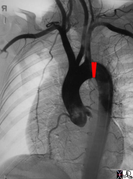
Isthmus
At the isthmus the aorta narrows by about 3mm. The isthmus defines the attachment of the ligamentum arteriosum to the aorta as well as delineating the arch from the descending aorta.
keywords aorta arch artery ascending descending normal thorax thoracic aorta ascending aortic valve sinus of Valsalva normal
Ashley Davidoff TheCommonVein.net 35324b04
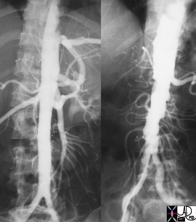
Abdominal Aorta
The abdominal aorta is a straight tube in the young and becomes elongated tortuous and enlarges with age
A Straight Tube but Gentle Narrowing of the Tubular Shape of the Aorta as it Proceeds from Proximal to Distal |
| 25661.800 aorta abdomen abdominal normal anatomy size celiac axis renal arteries Angiogram angiography Courtesy Ashley Davidoff MD |
Applied Biology
Thoracic Aorta
35290
Keywords aneurysm aorta artery pseudoaneurysm thorax trauma
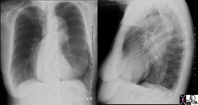
Arch
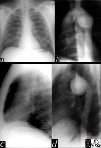
This image represents a combination of plain film CXR and the correlative thoracic aortogram in a 38 year old patient, 13 years after an MVA. There is an aneurysmal bulge at the level of the isthmus, representing a traumatic aneurysm at the characteristic location of the ligamentum arteriosum. The P-A and lateral chest X-ray shows an enlarged and unusually shaped aortic knob and the angiogram confirms the pseudoaneurysm of the aorta. 35178c Courtesy of Laura Feldman MD. TheCommonVein.net
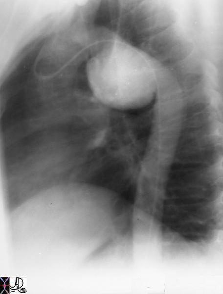
Saccular Pseudoaneurysm |
| This image represents an LAO view of the thoracic aorta in a 38 year old patient, 13 years after an MVA. There is an aneurysmal bulge at the level of the isthmus, representing a traumatic aneurysm at the characteristic location of the ligamentum arteriosum. 35178 Courtesy of Laura Feldman MD. code CVS aorta artery thorax trauma |
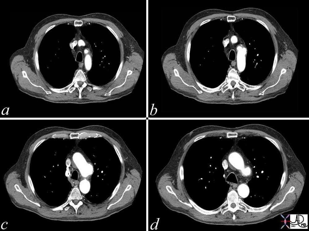
A series of CT scans showing a focal aneurysm of the aortic arch orignating just beyond the left subclavian artery. Whether this represents a focal saccular aneurysm from focal atherosclerotic weakening, or an ulcerating and penetrating plaque, or a site of previous dissection is difficult to know.
keywords code CVS chest thorax aneurysm aorta arch
Courtesy Ashley Davidoff MD. TheCommonVein.net 36817c
Takayasu’s Arteritis
Smooth Narrowing – Takayasu’s Aortitis |
| 20354b01 14 year old male artery thoracic aorta fx smooth narrowing of isthmus of aorta Takayasu’s aortitis angiography angiogram Courtesy Ashley Davidoff MD |
Descending
Tortuosity
Ectasia Elongation of the Aging Aorta resulting in a Sigmoid Shape |
| 71175c01 aorta thorax thoracic fx aortic ectasia sigmoid shape tortuosity breast asymmetry size CXR plain film Davidoff MD |
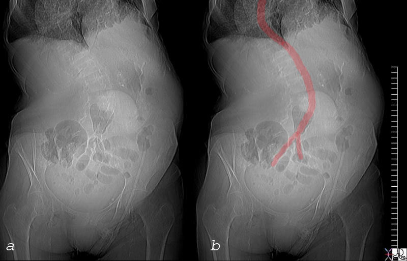
Abdominal Aorta
Atherosclerotic Disease
Tortuous Aorta – Normal Aging Process |
| The normal aging process results in a loss of elasticity. Elongation and tortuosity results which is exemplified in the image above, The patient is 60 years old
73384 Ashley Davidoff MD |
Aorta Follows the Shape of the Levoscopliosis |
|
Scoliosis associated with spina bifida 73838c01 bone lumbar spine shape aorta character levo scoliosis spina bifida CTscan scout view Courtesy Ashley Davidoff MD |
Normal and Severe Atherosclerosis |
| 18485c01 aorta artery hepatic artery renal artery splenic artery superior mesenteric artery SMA kidney severe atheroscleroris atheroma occluded renal artery spleen liver normal anatomy angiogram angiography Davidoff MD |
Abdominal Aortic Fusiform Aneurysm
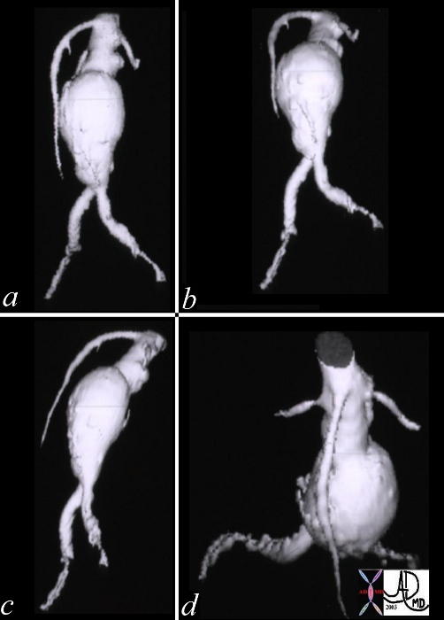
Fusiform AAA |
| This series of reformatted CT images showing a fusiform abdominal aortic aneurysm. The digital reconstruction enables the viewer to assess the aorta from multiple perspectives and the relationship of the aneurysm to the renal arteries and iliac arteries are easily assessed. These factors are important when surgical planning is considered. Angiography becomes unecessary when the detail of the aneurysm can be so exquisitely demonstrated. Courtesy GE Medical Sytems 10237c code aorta aneurysm infrarenal CTA AAA |
Smooth Narrowing
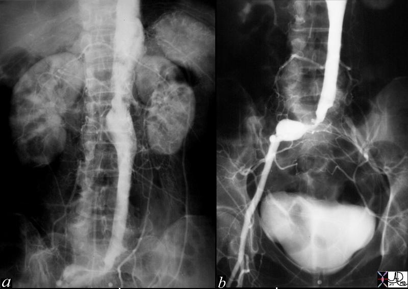
Smooth Narrowing of the Lumen Due to Aneurysmal Disease |
| This angiogram of the abdominal aorta (a) and iliac arteries (b), shows an unusually straight and narrowed infrarenal aorta indicative of thrombus in the wall of an abdominal aortic aneurysm. In addition there is an aneurysm of the right common iliac artery and a subtotal occlusion of the left common iliac artery. Note the left kidney is small and there is a wedge shaped defect in the upper and lateral aspect of the kidney indicative of an infarct, probably embolic in origin. Courtesy Laura Feldman MD. 36005c code abdominal aorta aneurysm artery iliac stenosis occlusion kidney infarct wedge embolus |
Inflammatory Disease
Smooth Narrowing
|
Takayasu’s Arterirtis |
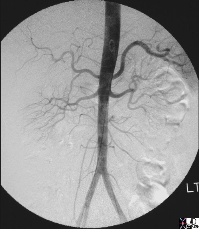
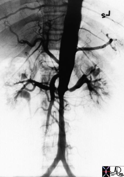
| The series of images are from the angiogram of a 14 year old female who presented with seizures and an elevated blood pressure. Images a and b show multiple stenoses within the carotids best seen at the level of the bifurcation into external and internal arteries. In addition in b, the aortic arch shows non critical narrowing just after the origin of the left common carotid vessel. Note that the right subclavian artery is not seen and presumably is occluded at its origin. The abdominal angiogram shows a significant narrowing of the left renal artery with post stenotic dilatation, and stenotic disease in the infrarenal abdominal aorta. The multicentric nature of the disease in a young female is pathognomonic of Takayasu’s arteritis. 35155c Courtesy of Laura Feldman MD. code CVS artery aorta arteritis inflammation Takayasu’s carotid thorax arch renal abdomen pulseless |
Irregular Narrowing
|
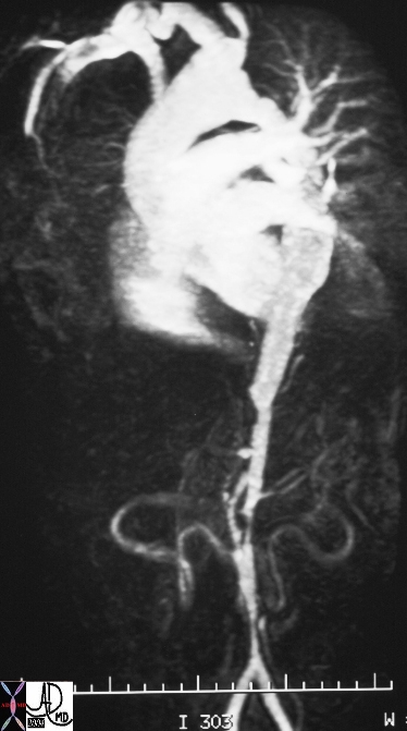
Takayasu’s Aortitis |
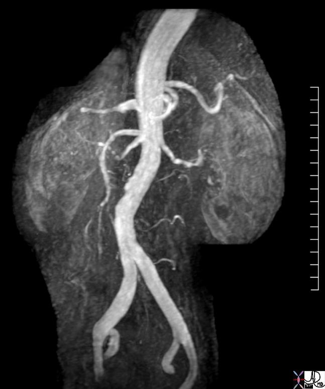
| 16917b aorta fx irregular fx narrow dx Takayasu arteritis imaging radiology MRI Courtesy Ashley Davidoff MD DB |
Focal Aneurysmal Disease
Mycotic Aneurysm
Mycotic Aneurysm |
| hx 68F with previous aortic surgery p/w back pain and fever aorta descending thoracic, thoracoabdominal fx periaortic fluid collection fx false aneurysm fx pseudoaneurysm fx paravertebral fluid collection bone vertebral body destruction dx mycotic aneurysm with dx vertebral osteomyelitis CTscan
Courtesy Ashley Davidoff MD 44502 |
Enlarging Pseudoaneurysm |
| 44514 hx 69M follow up July 2005 abdomen abdominal aorta fx pseudoaneurysm dx false aneurysm fx 6.6 by 6.2cms dx enlarging pseudoaneurysm CTscan Courtesy Ashley Davidoff MD |