
Copyright 2007
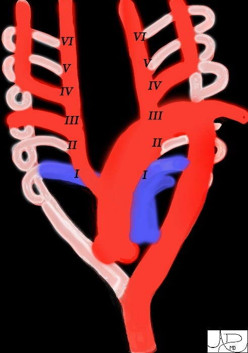
When pharyngeal arches are formed during the 4th and 5th weeks of development, each arch receives its own cranial nerve and its own artery. These arteries are known as aortic arches and arise from the aortic sac, the most distal part of the forming heart. The aortic arches terminate in the right and left dorsal aortae. In the region of the arches, the dorsal aortae remain paired, but inferior to this region they fuse to form a single dorsal aorta. There are 5 paired aortic arches numbered I, II, III, IV, and VI. In the 29 day embryo, aortic arches I and II have disappeared and III, IV, and VI are large and go on to form the great arteries (1) The 4th aortic arch persists on both sides, but its fate is different on the right and left. On the left, it forms part of the arch of the aorta, between the left common carotid and left subclavian arteries. On the right, it forms the most proximal segment of the right subclavian artery, the distal part of which is formed by a portion of the right dorsal aorta. (2) The distal portion of the right dorsal aorta disappears, and the left 4th aortic arch continuing into the left dorsal aorta and on to the common dorsal aorta, which is known as the abdominal aorta in adults
key words
artery embryology double arch left aortic arch segments 1 2 3 4 5 6 Davidoff Art Courtesy Ashley Davidoff MD TheCommonmvein.net 72814
Ashley Davidoff
Ashley Davidoff
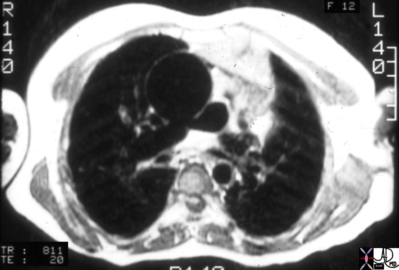
Key Words aorta pulmonary arteries branch pulmonary artery fx origin of the branch PA fromthe posterior aspect of the aorta dx TA truncus arteriosus MRI T1 weighted CHD Ashley Davidoff MD TheCommonmvein.net 01651.800