Iatrogenic Diseases of the Aorta
Copyright 2007
Ashley Davidoff MD

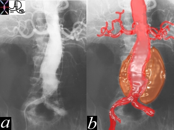
The Catheter Dissected through the Thrombus in the Aneurysmal Wall |
| This angiogram of the abdominal aorta shows a widened infrarenal aorta. At first glance the lumen of the aorta appears normal, but a faint curvilinar calcification of the true wall can be seen to the patients left in the first image. The second image (b) reveals the true size of the aneurysm. The key to the image though is to recognize that the catheter in image a, has left the contrast filled lumen and entred the thrombotic wall
Courtesy Ashley Davidoff MD 22734 cW02 codeCVS aorta artery abdomen aneurysm AAA |
