
Mural Hematoma
Copyright 2007
Ashley Davidoff MD
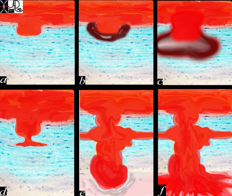
Pathogenesis of Penetrating Diseases of the Aorta and Acute Aortic Syndrome |
| 42409c01.800 aorta artery atherosclerosis atheroma acute aortic syndrome a aortic ulcer b = acute mural hematoma c = acute mural hematoma large d = focal dissection e penetrating ulcer f rupture histology histopathology Davidoff art pathogenesis Courtesy Ashley Davidoff MD |
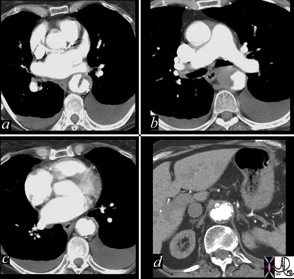
Mural Hematoma and Focal Dissection |
| This combination of images reflects the CTscan of a patient who presented with acute chest pain, and has findings reflecting a focal dissection of an atheromatous aorta. Note in (a) how thick the “intimal” flap is. Note also the mural dissection or hematoma with no flow within it. (b) Other parts of the descending aorta (c) and abdominal aorta (d) show only severe atheromatous disease. Courtesy Ashley Davidoff MD. 19416c code CVS aorta abdomen thorax dissection focal atherosclerosis |
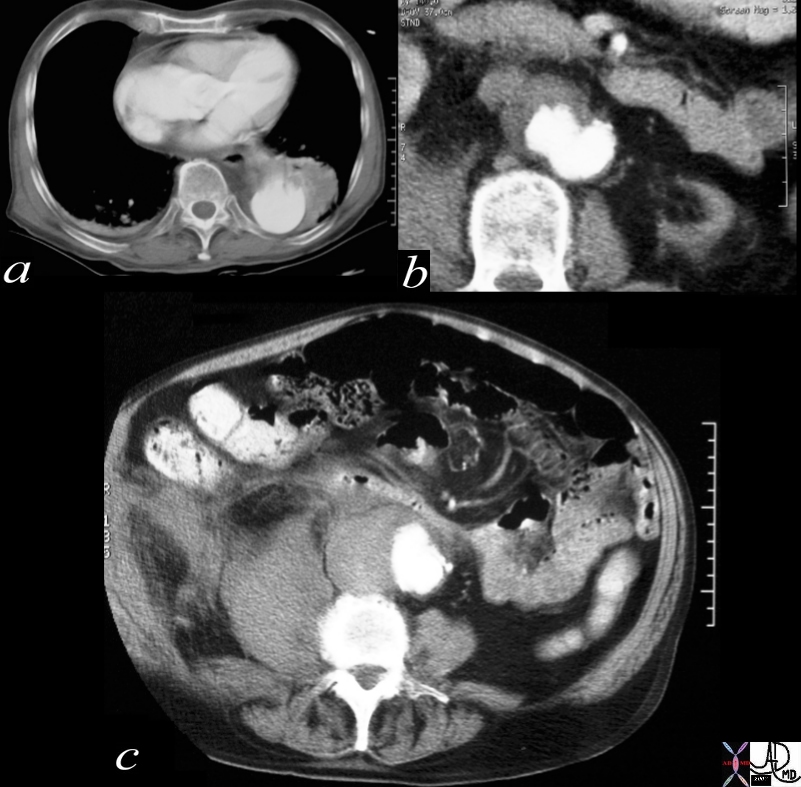
Penetrating Ulcer with Rupture |
| 17529c01 artery descending thoracic aorta abdominal aorta dx rupture pseudoaneumysm ulcerating plaque mural hematoma ruptured through aortic wall hemorrhage hematoma retroperitoneum CTscan Courtesy Ashley DAvidoff MD Ashley Davidoff MD |
Reference
Kang DH, Song JK, Song MG, Lee IS, Song H, Lee JW, Park SW, Kim YH, Lim TH, Park SJ. Clinical and echocardiographic outcomes of aortic intramural hemorrhage compared with acute aortic dissection.
Am J Cardiol. 1998 Jan 15;81(2):202-6.