
Aortic Aneurysm
The Common Vein Copyright 2007
Ashley Davidoff MD
An aortic anerusym is defined as an an enlargement of the aorta to a dimension greater than 1.5 times its normal diameter. Abdominal aneurysnmal disease is more common than thoracic aneurysmal disease.
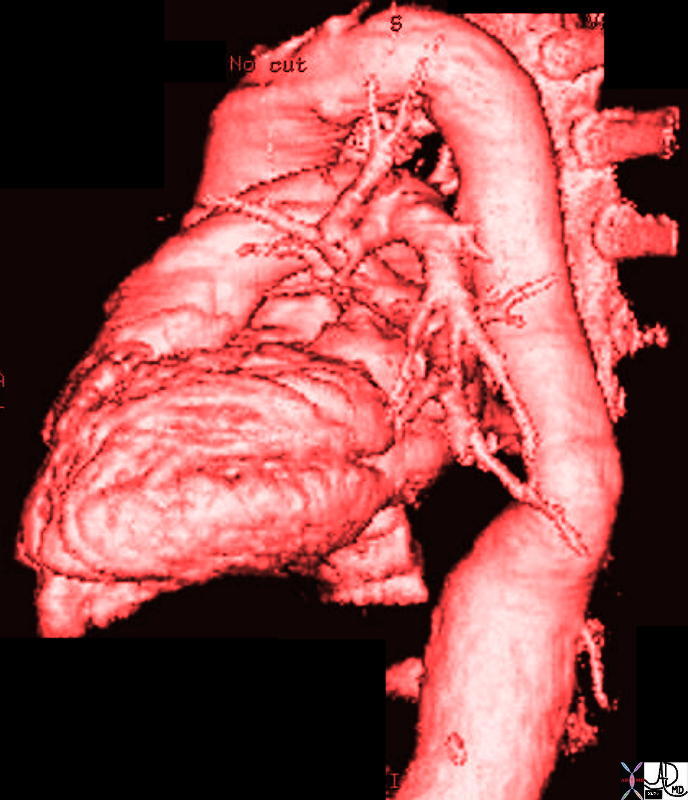 Suprarenal AAA Suprarenal AAA |
| 17061.801red aorta chest abdomen aneurysmal disease thoracic aorta abdominal aorta suprarenal aneurysm |
Cause
In the overwhelming majority the cause of aneurysmal aortic disease is related to the atherosclerotic process which causes weakening and loss of elasticity of the wall. In this disease the primary disease is in the intimal layer.Other diseases which accelerate the process in patients with atherosclerosis include hypertension, and hereditary aspects . Arteria magna is an entity of arteriomegaly with aneurysmal disease, usually associated with atherosclerosis and involves the aorta, iliacs, femoral and popliteal arteries. Medial degeneration is also an established cause of aneurysmal disease. In this disease there is degeneration of the media. It is a a feature of hereditary diseases such as Marfan’s syndrome and Ehlos- Danlos syndrome
Aortic infections may originate from the intima, within atherosclerotic plaque, from an external abscess, or via the vasa vasora. (such as occurs in syphilitic disease).
Age
Patients are more commonly males between 60 and 70 years.
Statistics
95% of aneurysms involkve the infrarenal artery. 30% of patients with aneurysms smaller than 6cms ruptured. (Szilagyi) 40% of patients with aneurysms greater than 6cms rupture
Anatomic Considerations:
The aorta is mostly an elastic artery so that it can act as a secondary “pump” to propel the blood to the furthermost structure. The infrarenal aorta has fewer vasa vasora than the thoracic aorta. This theoretically renders it more “ischemic”
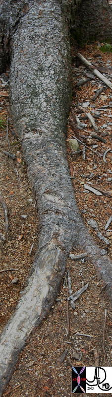 
“Aneurysms” in Nature |
| This image of an above ground root taken in Black Cap mountain near Conway NH, shows aneurysmal dilatation of the proximal part of the root.
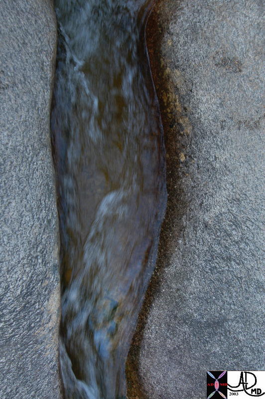
The second image of a rivulet through a crack in the rock at Diana’s baths in Bartlett NH, shows mild aneurysmal dilatation of the proximal portion of the crack reminiscent of an infrarenal AAA. Note the non-laminar flow in the downstream portion of the flow as the diameter of the rivulet decreases. Courtesy Ashley Davidoff MD 73553p code tree aneurysm AAA root interesting accessory Courtesy Ashley Davidoff MD 73953p code accessory interesting rock aneurysm water |
Classification
Based on site
Aneurysm are classified based on theitr location in the aorta, as ascending arch, descending thoracic, thoracoabdominal, suprarenal, or infrarenal.
Based on wall structure
Aneurysm are true aneurysm when all three layers are part of the wall, whereas a false aneurysm is characterised by the absence of vessel wall, and the wall of the aneurysm is created by surrounding thrombus, and is contained by the thrombus and surrounding tissues.
Based on shape
A fusiform aneurysm is a spindle-shaped aneurysm implying that it is widest near the middle and tapers in toward both ends. Atherosclerotic aneurysms are classically fusoiform. A saccular aneurysm is more focallly eccentric and round. Mycotic aneurysms are calssically saccular. Saccular aneurysm tend to have a higher propensity for rupture. They occur between fixation points where there is focal loss of elastin and collagen and an increase in elastase activity
Pathogenesis
In atherosclerosis disease starts in the intima with secondary effects on the media. Once the medial elements are destroyed. there is weakening of the wall, and the vessel dilates. As the tension increases, Laplace’s Law (tension is proportional to the product of pressure and radius) dictates that the tension is greatest on the largest diameter and the tension is therefore exertd on the weakest part of the wall. Progressive dilatation of the wall therefore occurs
Historical Aspects
In the second century AD Galen described a ruptured aneurysm – “bright red blood spurted forth with much violence.” The first successful surgical repair of an aneurysm was in 1955. About 3/4 of atherosclerotic aneurysms are confined to the abdominal aorta Most occur between the renal arteries and aortic bifurcation Only 2-5% are suprarenal and these are usually extensions of thoracic aneurysms Abdominal aneurysms are usually fusiform and may contain laminated thrombotic material due to stagnant flow
Natural History
The normal aorta grows in diameter by 1mm per year. An established aneurysm will grow by 2-4mm per year. Half of aneurysms greater than 6 cm will rupture within a year 25% of aneurysms 4-6 cm will rupture in a year 15-20% of smaller aneurysms will rupture in a year The average aneurysm expansion rate is 0.4 to 0.5 cm per year but increases as the aneurysm increases in size
Diagnosis
Clinical
esults in a pulsating feeling and abdominal and back pain, coughing, wheezing, Horner’s syndrome, hoarseness, and swallowing difficulty.
Diagnostic Tools
Best accomplished by cross-sectional ultrasound (simple and accurate) Aortic angiography may not demonstrate the full width due to nonopacified thrombus Angiography is indicated If there is a question of the correct diagnosis For hypertensive patients with possible renal artery disease (to confirm or exclude renal artery stenosis) To determine definitively the extent of involvement CT and MRI are also excellent for diagnosis and sizing of larger aneurysms
Treatment
Elective surgery of all abdominal aneurysms is recommended when they exceed 5cms. cm Management of asymptomatic aneurysms of <5 cm is controversial and close follow-up of poor surgical candidates with aneurysms of 4-6 cm is advisable Late survival is not affected by surgery with 50% 5 year survival of patients with unresected aneurysms <6 cm compared to 60-70% survival with resection Operative mortality is 2-5% for elective resection in low-risk patients, 5-15% for expanding aneurysms and 50% with rupture The operative mortality for elective surgery is 2-3%. In the situation where there is artic rupture, the mortality for surgery is 50-90%. Treated by surgery.
References
CT signs of pending Aortic aneurysm rupture web presentation from radiology assistant .nl 5*
Gonsalves crescent sign
Pillari letter to the editor