
Lecture Big Red 05 25 21
Acute Aortic Syndrome 7/6/21
Case Presentations 7/8/21
Ashley Davidoff
The Aorta

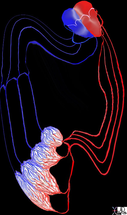
The heart and lungs are positioned at the centre of all the organs in the body and they are understandably considered vital organs. Think of yourself as a little red cell within the vast and powerful environment of the circulatory system. Listen and feel the power of the pulse and the rush of all the rivers going back and forth, hither and thither, and around and around – imagine what goes on inside the system all the time – it is probably like walking through a rain forest with huge waterfalls, winding rivers and rivulets – a mixture of gushing power and calm meandering flow- but the colors are reds and blues instead of greens and yellows! Courtesy Ashley Davidoff MD 32059

The heart and lungs are positioned at the centre of all the organs in the body and they are understandably considered vital organs. Think of yourself as a little red cell within the vast and powerful environment of the circulatory system. Listen and feel the power of the pulse and the rush of all the rivers going back and forth, hither and thither, and around and around – imagine what goes on inside the system all the time – it is probably like walking through a rain forest with huge waterfalls, winding rivers and rivulets – a mixture of gushing power and calm meandering flow- but the colors are reds and blues instead of greens and yellows!
Ashley Davidoff MD 32059d01 key words cardiac heart circulation artery vein introduction drawing Davidoff art
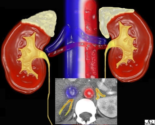
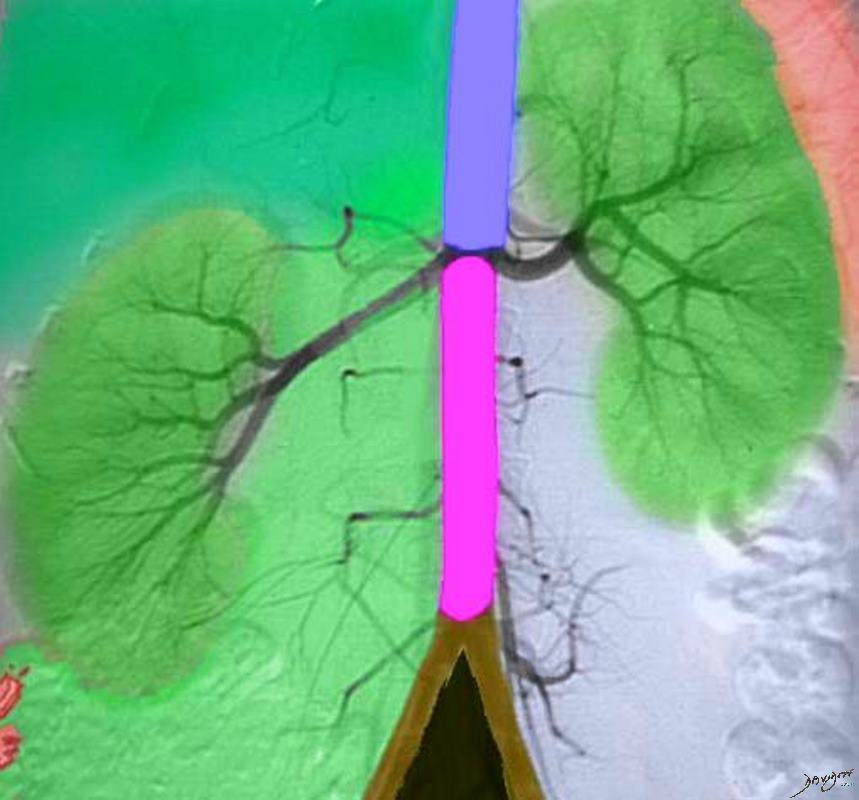
39533 This image combines the coronal view with the axial view and reflects the intimate relationships that the adrenals have with the kidneys as well as the great vessels of the abdomen. They literally have their fingers on the pulse of the aorta (red overlay)and the inferior vena cava (IVC) (blue overlay)
Ashley Davidoff
keywords
adrenal kidney relations anatomy
32368b05.800
Ashley Davidoff MD

72845.800 aorta flow principles structure laminar flow turbulent flow normal
Ashley Davidoff MD 72835.800 72839 72831.800 72845.800 49483b01
72831.800 aorta flow principles structure laminar flow turbulent flow normal tube Davidoff Art Courtesy Ashley Davidoff MD 72835.800 72839 72831.800 72845.800 49483
49483b01
key words
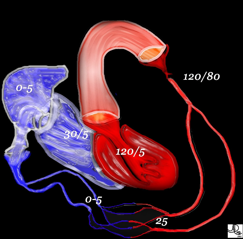
heart cardiac LV left ventricle aorta aortic systemic circulation capillary capillaries arterioles venules right atrium RA right ventricle RV normal physiology pressures hemodynamics
Ashley Davidoff
49483b01 heart cardiac LV left ventricle aorta aortic systemic circulation capillary capillaries arterioles venules right atrium RA right ventricle RV normal physiology pressures hemodynamics Ashley Davidoff

This diagram reflects the laminar flow showing the almost parallel lines of flow that is characteristic of flow in the smaller tubes. Under resting conditions, laminar flow exists from the medium-sized bronchi onward down to the bronchioles. During exercise, the air flow is accelerated, and laminar flow may be confined only to the very small airways.
Ashley Davidoff MD 42433b04.jpg
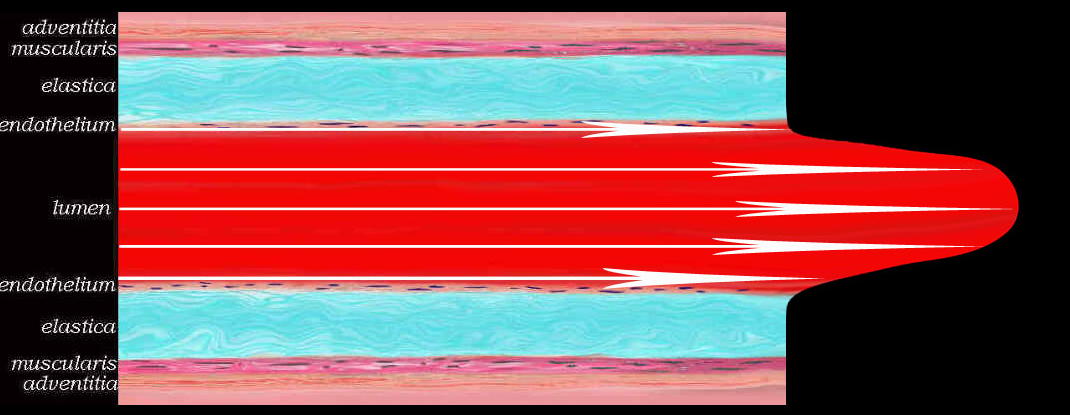
keywords velocity parabolic Laminar flow is the normal condition for blood flow throughout most of the circulatory system. characterized by concentric layers of blood move in parallel down the length of a blood vessel. highest velocity (Vmax) isin the center of the vessel. lowest velocity (V=0) is along the vessel wall. T flow profile is parabolic occurs in long, straight blood vessels, under steady flow conditions. Davidoff art Courtesy Ashley Davidoff MD
47678d05
keywords velocity parabolic Laminar flow is the normal condition for blood flow throughout most of the circulatory system. characterized by concentric layers of blood move in parallel down the length of a blood vessel. highest velocity (Vmax) isin the center of the vessel. lowest velocity (V=0) is along the vessel wall. T flow profile is parabolic occurs in long, straight blood vessels, under steady flow conditions. Davidoff art Courtesy Ashley Davidoff MD
47678f12.800

This drawing shows the lines and circles of noisy turbulent flow, which is characteristic of flow in the larger airways.
Courtesy of Ashley Davidoff M.D. 42434b07
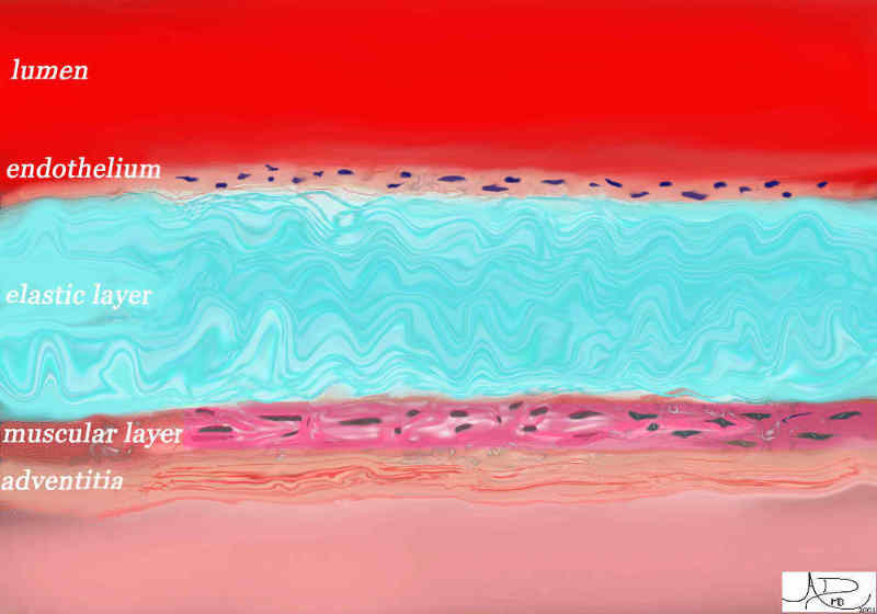
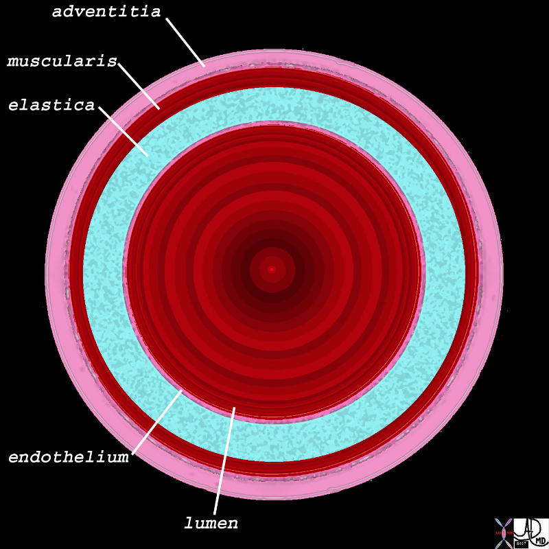
The Innermost Layer is the Endothelium and Outermost is the Adventitia
47678.800
Ashley Davidoff MD

The Innermost Layer is the Endothelium and Outermost is the Adventitia
47678.800 Ashley Davidoff MD
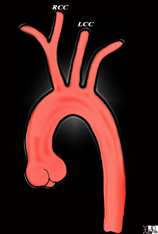
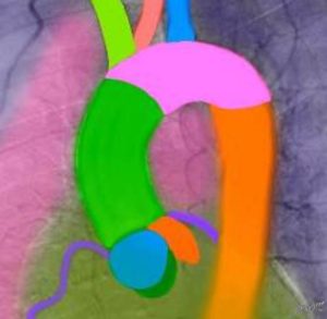
keywords aorta aortic valve sinotubular junction ascending aorta aortic arch brachiocephalic artery left common carotid artery left subclavian artery descending aorta normal TCV
Ashley Davidoff MD
47676
Key Words
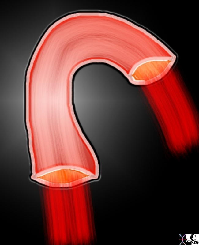
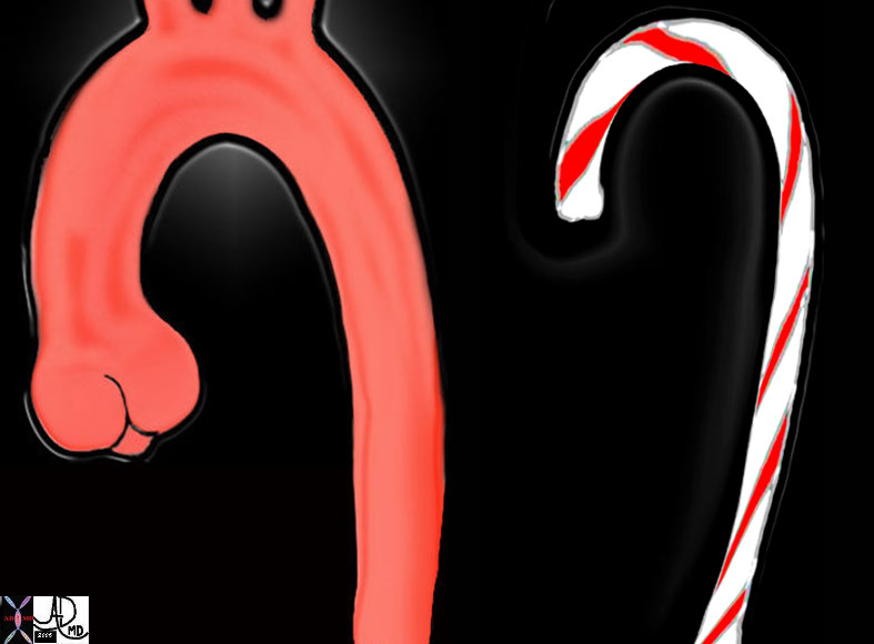
candy cane shape aorta aortic valve sino-tubular junction ascending aorta aortic arch descending aorta normal
Ashley Davidoff
47677c03

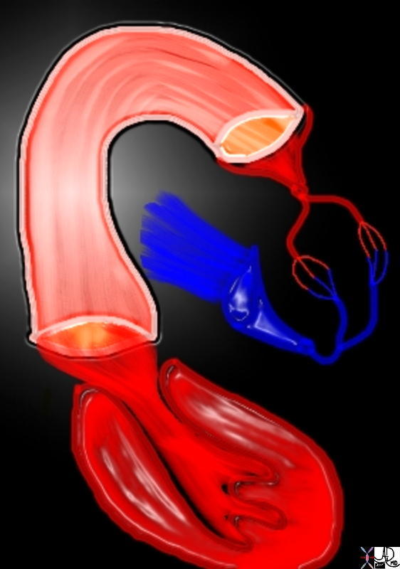
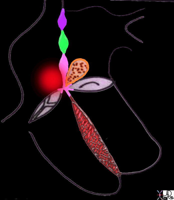
The aortic valve (AV) lies central to many structures including the right ventricular outflow tract (RVOT), pulmonary valve (PV), left atrium (LA), right atrium (RA), and superior vena cava (SVC). aorta from above. In this diagram, the commissures are overlaid in green, the crescent shaped lunulae reflect the free edge of the leaflets and are overlaid in yellow, and the nodules of Arantii are overlaid in orange. The sinuses of Valsalva are like cups and are positioned between the free edges. (gray)
47681d07
Ashley Davidoff MD
01669b04
Ashley Davidoff MD 01667b04 01667b04 01667b06 01667b07 01667b09 01667b14 01669b03