
Structure
The aorta as part of the cardiovascular system is the largest and most integral artery of the circulatory system. It’s structure is characterized by a tubular form and elastic properties. Arising from the left ventricle of the heart, the aorta’s overall structure is divided into three sections: the ascending aorta, arch of the aorta and the descending aorta. The descending aorta has a thoracic portion which enters the abdominal cavity through the aortic hiatus to become the abdominal aorta which terminates at the iliac bifurcation.
The aorta is histologically comprised of three layers: the intima, the media and the adventitia. Such a composition is characteristic of its function as a distensible tube which supplies oxygenated blood to the heart and body.
|
Function
Its sole function is to deliver oxygenated blood from the heart to the first order branches of arteries at a sufficiently high pressure so that they can sequentially transport the oxygenated blood to the capillary system and then on to the tissues. Its functional reach therefore is to all organs and structures of the body through arteries, arterioles and capillaries.
The branches
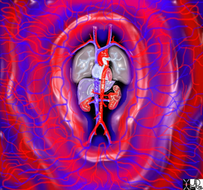
The branches of the aorta get progressively smaller as they divide until the capillary system is reached. Flow into the capillary system is controlled by the precapillary sphincters. As blood flows through the capillaries, fluid rich in oxygen, is exchanged for fluid that is rich in waste products. This fluid is removed and returned to the heart via the venous system.
 End Point of the Arterial Circulation is at the Capillaries and is also the Beginning of the Venous Circulation End Point of the Arterial Circulation is at the Capillaries and is also the Beginning of the Venous Circulation |
| 32368b05.800 cardiovascular system heart cardiac artery arteries capillary capillaries arterioles venules veins negative firces positive forces road highway TCV the common vein applied biology Davidoff art Davidoff MD |
Diseases
The most common disease of the aorta is atherosclerosis. It is a degenerative disease which normally occurs with aging. The atherosclerotic process can be accelerated and advanced in patients with genetic predisposition, dietary indiscretion or cigarette smoking. This accelerated degeneration may result in aneurysmal disease. Other less common diseases of the aorta include aortic dissection and aortitis.
The Normal and Atherosclerosis of the Aorta |
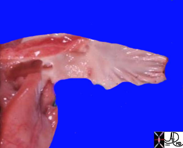
| The first image shows the aorta of a baby(light pink color) coming from the heart (red) and it is characterised by its smooth and glistening nature. The second image shows a moderate amount of atherosclerosis characterized by irregular yellow fibrofatty nodules on the otherwise smooth and glistening surface of the endothelium or inner lining of the aorta.
Courtesy Ashley Davidoff 0157b Courtesy Henri Cuenoud MD 13420c 13418 13419 13420 |
Aneurysms are another common disease that affect the aorta caused by loss of elasticity of the wall and almost always associated with atherosclerosis. There are a few types of aneurysms that are caused for example by trauma and infection that are not associated with atherosclerosis.
 The Normal and Aneurysmal Disease The Normal and Aneurysmal Disease |
| This image shows a moderate amount of atherosclerosis characterized by irregular yellow fibrofatty nodules on the otherwise smooth and glistening surface of the endothelium or inner lining of the aorta.
Courtesy Henri Cuenoud MD 13420c 13418 13419 13420 |
Diagnosis
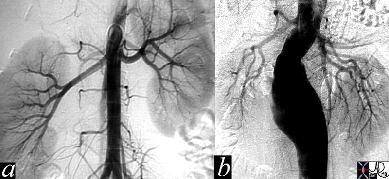
Current diagnostic procedures for aortic disease utilize CT scan and MRI most commonly. Transesophageal echocardiography is reserved for evaluation of the thoracic aorta and ultrasonography for the abdominal aorta with the latter being particularly useful for the evaluation of abdominal aortic aneurysms. Angiography in the past was the gold standard for both thoracic and abdominal evaluation, but because of its relative invasiveness, and the advancing capabilities of CT scan and MRI, it has become a second line study.
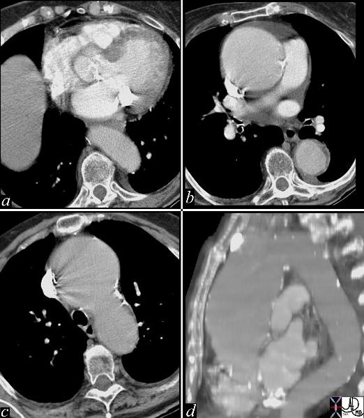 CT scan of an aneurysm of the Ascending Aorta CT scan of an aneurysm of the Ascending Aorta |
| This series of images are from a CTscan showing an ascending aortic thoracic aneurysm. There is evidence of heavy calcification of the aortic valve (aortic sclerosis), an aneurysm confined to the ascending aorta (b,c,d), and tortuosity of the descending aorta (d). The cause for the aneurysm is probably a combination of systemic hypertension, aortic stenosis and atherosclerotic degeneration of the wall.
Courtesy Ashley Davidoff MD 19426c code CVS thorax aorta ascending aneurysm MAC aortic sclerosis CTscan |