|
Imaging Aneurysmal Disease
The Common Vein Copyright 2007
Sinus of Valsalva
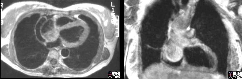
Ascending Aorta
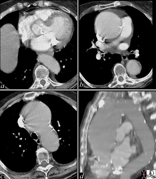
Ascending Aortic Aneurysm
|
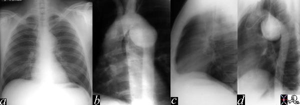
| This series of images are from a CTscan showing an ascending aortic thoracic aneurysm. There is evidence of heavy calcification of the aortic valve (aortic sclerosis), an aneurysm confined to the ascending aorta (b,c,d), and tortuosity of the descending aorta (d). The cause for the aneurysm is probably a combination of systemic hypertension, aortic stenosis and atherosclerotic degeneration of the wall. Courtesy Ashley Davidoff MD 19426c code CVS thorax aorta ascending aneurysm MAC aortic sclerosis CTscan |
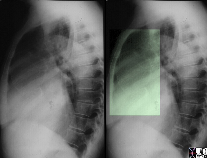
Syphilitic Aortitis
|
| This lateral examination of the chest shows fine calcification in an ectatic ascending aorta associated with aortic annular calcification. Note the remarkable paucity of atherosclerotic change in the descending aorta. These findings are highly characteristic of tertiary syphilis of the aorta. Courtesy Ashley Davidoff MD. 00018c code CVS artery aorta ascending syphilis aneurysm calcification tortoise shell |
Arch
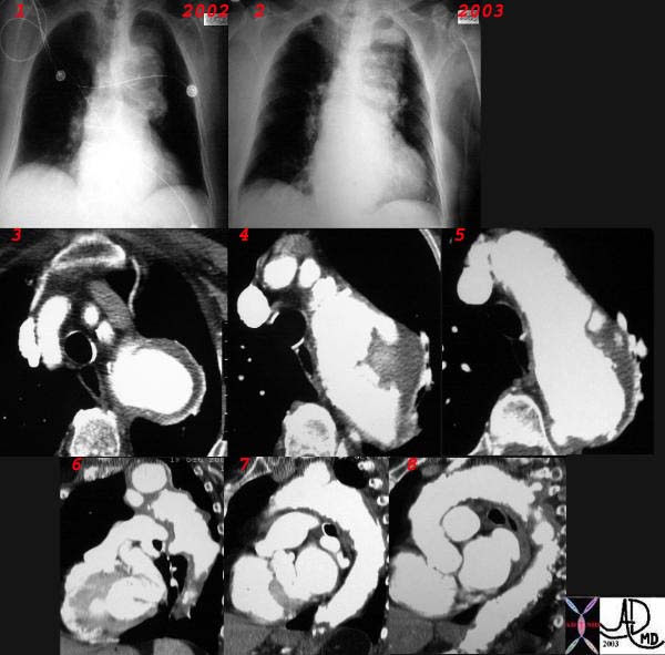
Arch Aneurysm
|
| This combination CXR and CT reveals an expanding aneurysm of the arch fromm 2002 to 3003. The CT shows three aneurysm in the arch of the aorta. The largest seen in image 3,6, and 7 accounts for the finding in the left apex of the CXR, while a second pseudoaneurysm is seen on the lateral border of the knob (4,8) and a penetrating ulcer medially (5) 32029c |
Traumatic Pseudoaneurysm
|
| This image represents a combination of plain film CXR and the correlative thoracic aortogram in a 38 year old patient, 13 years after an MVA. There is an aneurysmal bulge at the level of the isthmus, representing a traumatic aneurysm at the characteristic location of the ligamentum arteriosum. The P-A and lateral chest X-ray shows an enlarged and unusually shaped aortic knob and the angiogram confirms the pseudoaneurysm of the aorta. 35178c Courtesy of Laura Feldman MD. code CVS aorta artery |
Descending Thoracic Aorta
Abdominal Aorta
AAA
|
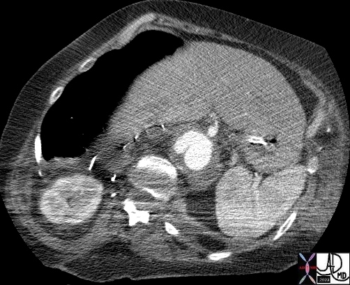
| 17061.801red aorta chest abdomen aneurysmal disease thoracic aorta abdominal aorta suprarenal aneurysm |
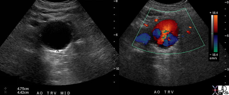
Turbulence in an Abdominal Aortic Aneurysm
|
| 37669c01 aorta abdomen abdominal aort AAA enlarged aneurysm abdominal aortic aneurysm turbulent flow USscan color flow doppler Courtesy Ashley Davidoff MD |
Smooth Narrowing of the Lumen Due to Aneurysmal Disease
|
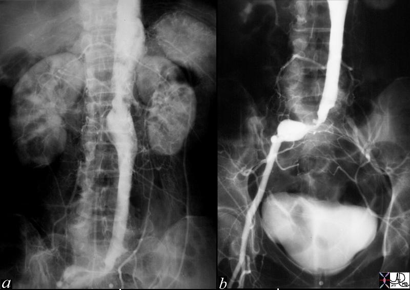
| This angiogram of the abdominal aorta (a) and iliac arteries (b), shows an unusually straight and narrowed infrarenal aorta indicative of thrombus in the wall of an abdominal aortic aneurysm. In addition there is an aneurysm of the right common iliac artery and a subtotal occlusion of the left common iliac artery. Note the left kidney is small and there is a wedge shaped defect in the upper and lateral aspect of the kidney indicative of an infarct, probably embolic in origin. Courtesy Laura Feldman MD. 36005c code abdominal aorta aneurysm artery iliac stenosis occlusion kidney infarct wedge embolus |
Mycotic Aneurysm
|
| hx 68F with previous aortic surgery p/w back pain and fever aorta descending thoracic, thoracoabdominal fx periaortic fluid collection fx false aneurysm fx pseudoaneurysm fx paravertebral fluid collection bone vertebral body destruction dx mycotic aneurym with dx vertebral osteomyelitis CTscan
Courtesy Ashley Davidoff MD 44502 |
AAA CT Angiogram
|
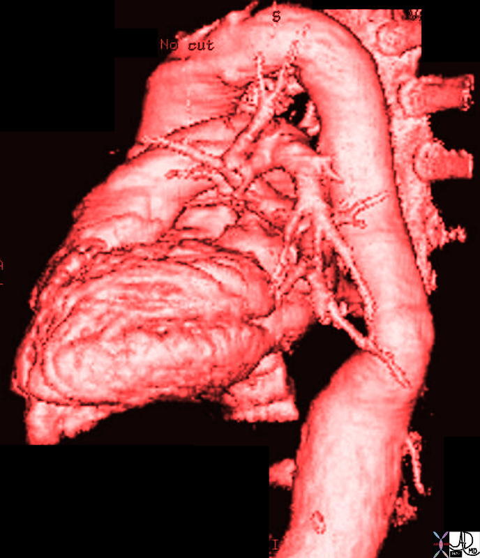
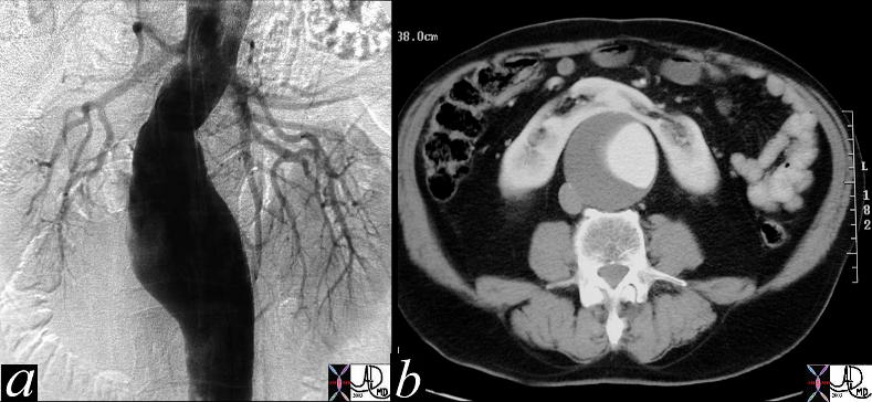
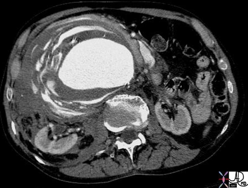
| This digital angiogram of an infrarenal, fusiform abdominal aortic aneurysm, shows an expanded and tortuous aortic lumen. The contrast column represents the luminal diameter and not the true size of the aneurysm. The size is best evaluated on cross sectional imaging where the outside edges of the wall are seen to best advantage. (b). Note there is a large thrombotic component only appreciated on the CT scan. A horseshoe kidney is vaguely outlined on the angiogram, but is quite obvious on the CT scan. This is an essential piece of information for the surgeon, and makes the surgery that much more difficult. Courtesy Ashley Davidoff 11976c code CVS aorta abdomen AAA kidney horseshoe |
Crescent Sign
|
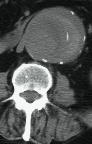
| 25253b abdomen aorta abdominal aortic aneurysm AAA fx crescent sign fx hyperdense rim fx calcificatoion in thrombus dx impending rupture imaging radiology CT scan Courtesy Ashley Davidoff MD |
Crescent Sign and Rupture
|
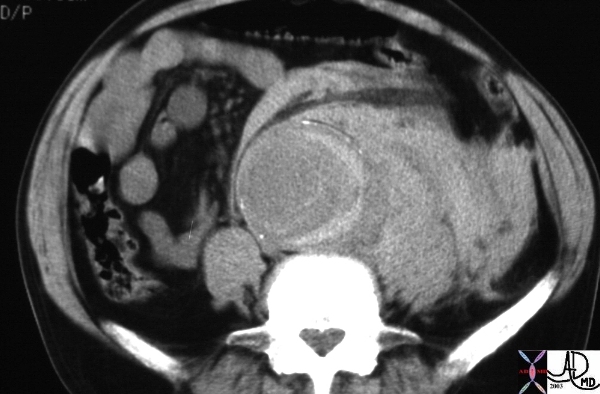
| The CTscan of the abdominal aorta reveals an acute rupture characterised by a high density crescent shaped density of acute blood within the chronic thrombus of the aortic lumen. The extraluminal component of the rupture is seen as high density acute blood in the retroperitoneum. Courtesy Ashley Davidoff MD 27175 code abdomen aorta hemorrhage retroperitoneum fx crescent sign dx abdominal aortic aneurysm AAA rupture |
Ruptured AAA
|
| 18269 aorta abdomen AAA aortic aneurysm abdominal aorta retroperitoneum fx retroperitoneal hematoma fx active hemorrhage fx perinephric hematoma anterior pararenal space perirenal space posterior pararenal space hemorrhage dx rupture abdominal aortic aneurysm CTscan Davidoff MD fx ruptured AAA |
|
