
MRI
Copyright 2007
Outflow Level
Aortic Valve
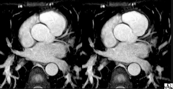
Normal Aortic Valve Cusps and Left Coronary Artery |
| This axial MRI view is shows the RVOT anterior and leftward of the aortic valve, after it has twisted around the aorta. The thin musculature of the ovoid infundibulum is in red overlay in the second image. Courtesy of Philips Medical Systems and modified by Ashley Davidoff M.D.) 32092 |
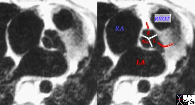
Valve Leaflets T1 sequence |
| 07954bWduo heart cardiac aorta aortic valve infundibulum RVOT right ventricular outflow tract position relation MRIscan Davidoff MD |
D Transposition |
| 07258b02 anterior aorta posterior pulmonary artery position connection subaortic conus D TGA TGV D transposition of the great vessels D transposition of the great arteries dextro rightward MRI Davidoff MD |
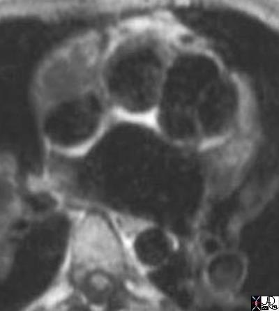
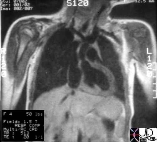
Sinus of Valsalva Aneurysm |
| 07974c01 heart cardiac right atrium aortic valve aortic sinus fx right atrial filling defect dx sinus of Valsalva aneurysm of the right aortic sinusprolapsing into the right atrium (RA defect) dx Marfan’s Syndrome MRI T1 weighted Davidoff MD 07973.800 07974c01 07974.800 |
Supravalvar Aortic Stenosis |
| Hx 4 year female with cocktail pesonality hearyt cardiac artery aorta supravalvular aortic stenosis supravalvar aortic stenosis Williams syndrome William’s syndrome
08233b01 |
Truncus Arteriosus |
| 01651.800 aorta pulmonary arteries branch pulmonary artery fx origin of the branch PA fromthe posterior aspect of the aorta dx TA truncus arteriosus MRI T1 weighted CHD |
Isthmus

Coarctation |
| This combination of an angiogram and MRI show a focal coarctation of the aorta distal to the dilated left subclavian artery. Note the large internal mmammary artery in the angiogram (a) Courtesy Ashley Davidoff MD 00254c02 code CVS aorta thorax coarctation |
Arch and Descending
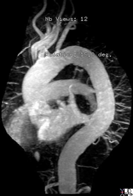
Tortuos Thoracic Aorta |
| 38852.800 thorax abdomen ascending aorta aortic arch descending thoracic aorta abdominal aorta shape tortuous MRI Courtesy Ashley Davidoff MD |
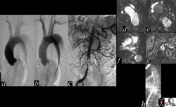
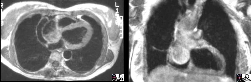
Aortic Dissection – Angiography and MRI |
| The thoracic angiogram (a,b)reveals an aortic dissection which starts just after the left subclavian artery. The true lumen is the narrowed medial lumen which is denser than the larger lateral false lumen. In (c) the A-P view reveals a patent proximal abdominal aorta, but the distal narrowing and occlusion of the the left iliac artery suggests that the dissection has progressed downstream. In the cross sectional MRI images (d,e,f,g) the intimal flap separating the true and false lumena is quite obvious. The true lumen is medial and narrow while the false is lateral and larger. Note that the SMA in g, arises off the smaller true lumen. In image h, a coronal view of the aorta, reveals the true narrow lumen anteriorly with faster (more intense) flow, and the posterior larger lumen with slower flow. Courtesy Laura Feldman MD. 35124c code CVS artery aorta dissection thorax abdomen angiography MRI |
Abdominal Aorta
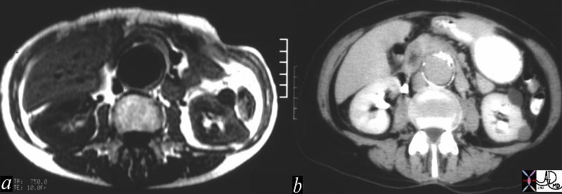
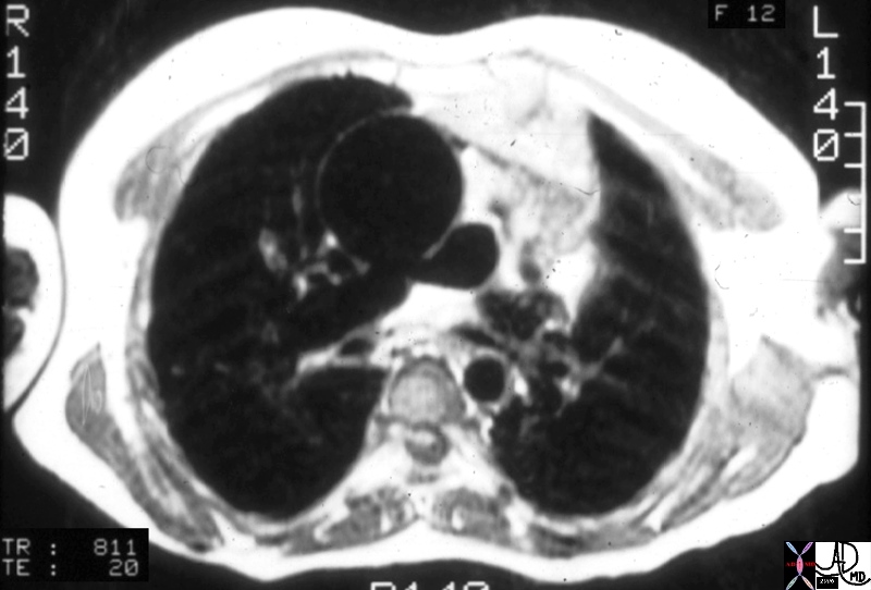
MRI vs CT for AAA |
| These two images represent cross sectional imaging of an infrarenal fusiform abdominal aortic aneurysm, with image a, being from a T1 weighted MRI sequence and image b from a CT scan with contrast. MRI fails to have the sensitivity for calcium that CT does, and since calcification is an intrinsic part of aneurysmal disease, when it comes to meausurement of aortic aneurysmal size, CT is superior. Courtesy Ashley Davidoff MD 15153c02 code CVS aorta abdomen AAA aneurysm fusiform infrarenal |
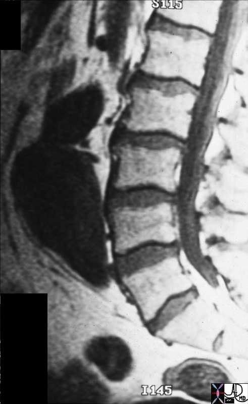
AAA |
| 23263b back pain aorta artery abdominal aortic aneursym fusiform shape spine bone MRI T1 sagittal Courtesy James Donnelly MD |
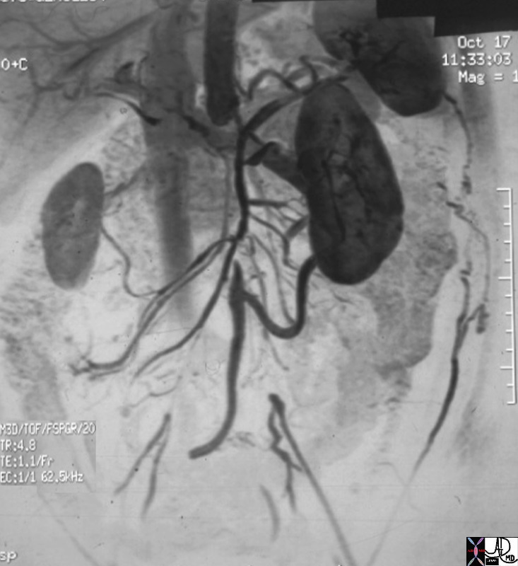
Aortic Occlusion |
| 14646.800 aorta kidney artery superior mesenteric artery arc of Drummond IMA inferior mesenteric artery collaterals renovascular disease occluded aorta internal iliac artery fx occluded occlusion small compensatory hypertrophy collaterals MRA MRIscan Davidoff MD |
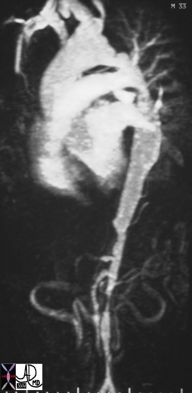
Takayasu’s Arteritis |
| 16910b aorta thorax fx irregular fx narrow dx Takayasu arteritis imaging radiology MRI Courtesy Ashley Davidoff MD DB |