Copyright 2007
Annulus
Calcified Annulus and Calcified Atherosclerotic Ascending Aorta |
| 72859c01 aorta aortic annulus fx calcified calcification dx aortic sclerosis CTscan Courtesy Ashley Davidoff MD |
Aortic Valve
Normal Aortic Valve |
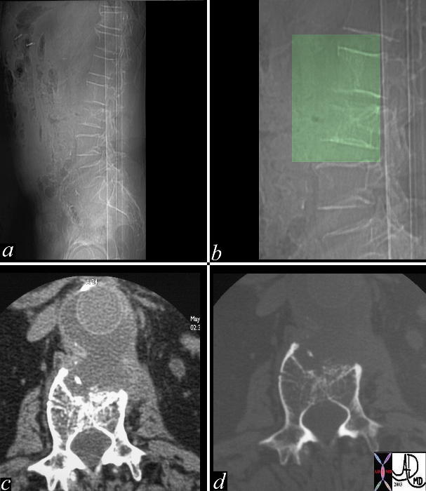
| 47368c02 spine bone thoracic spine ospeopenia osteoporosis fx wedge compression fractures shape mechanical forces calcification aortic valve aortic sclerosis aortic stenosis AS kyphosis CXR plain film X-ray Davidoff MD |
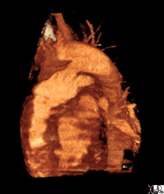
Bicuspid Aortic Valve |
| 72885.800 thorax thoracic aorta aortic valve fx thickened dx bicuspid aortic valve CTscan Courtesy Ashley Davidoff MD |
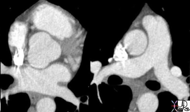
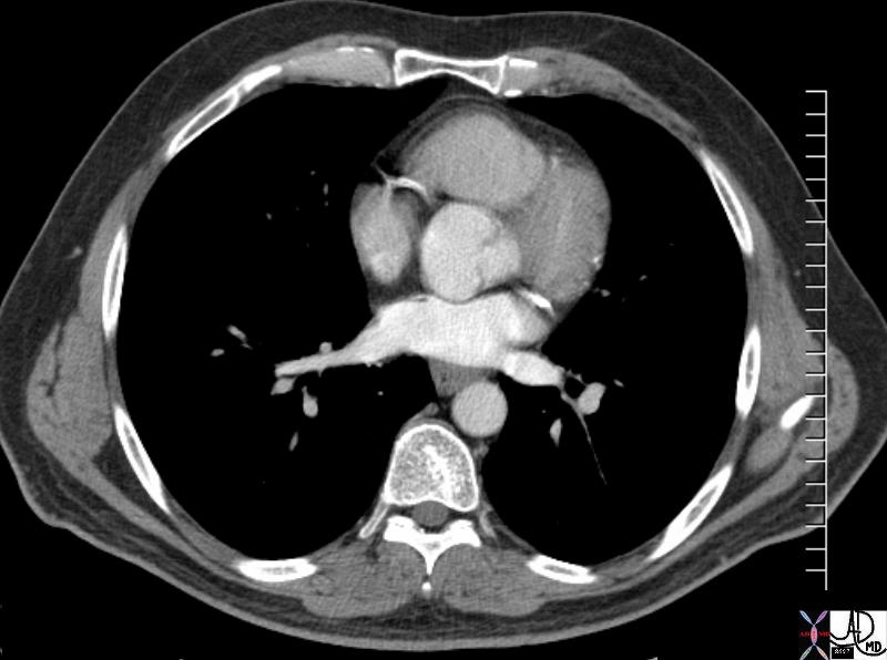
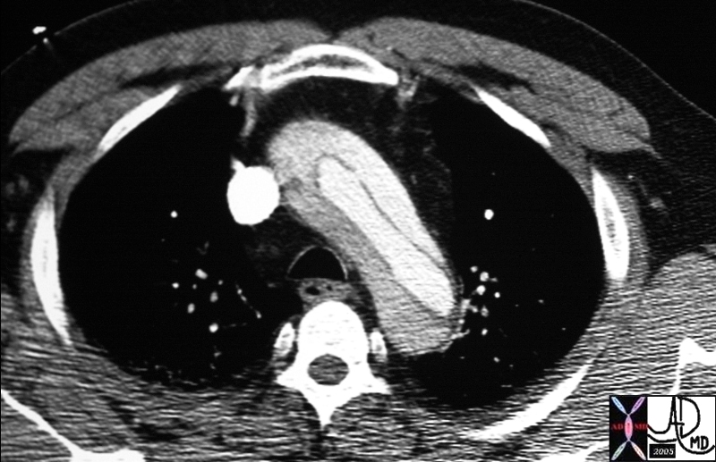
Calcific Aortic Stenosis |
| 39698 heart cardiac aortic valve calcification aortic stenosis LVH |
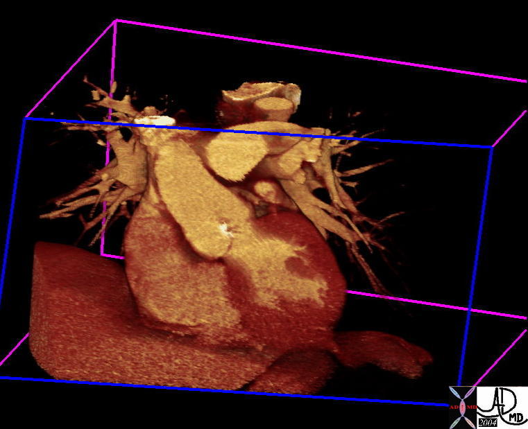
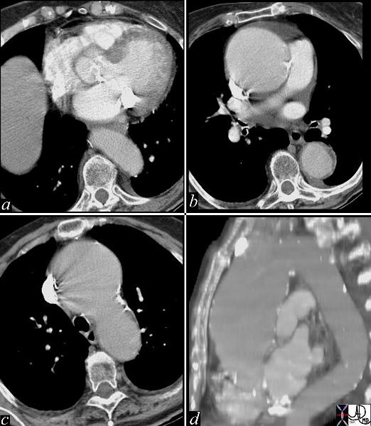
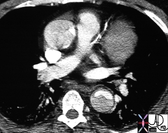
Calcific Aortic Stenosis with Post Stenotic Dilatation |
| 45196 heart cardiac RA right atrium right ventricle RV RVIT RVOT pulmonary valve TV tricuspid valveright ventricular sinus right ventricular outflow tract aorta aortic calcification calcified aortic valve dx AS aortic stenosis anatomy CTscan Courtesy Ashley Davidoff MD |
Ascending Aorta
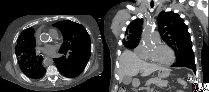
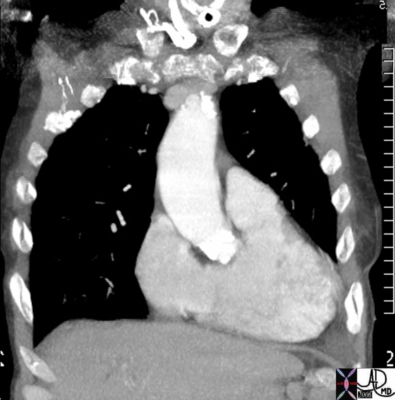
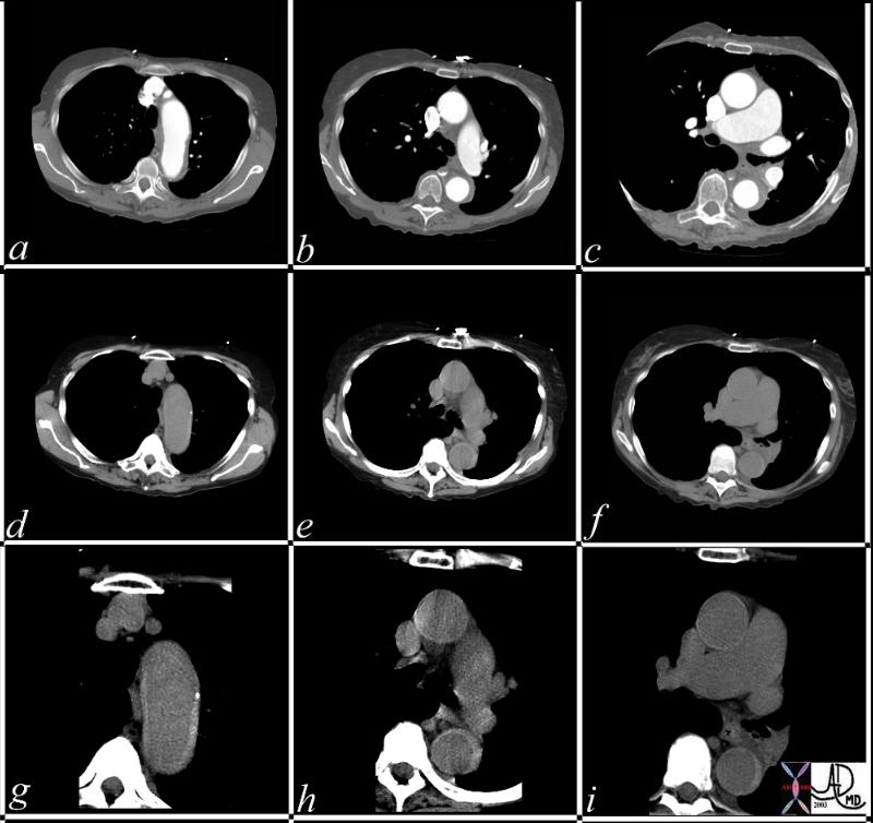
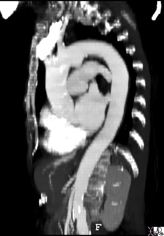
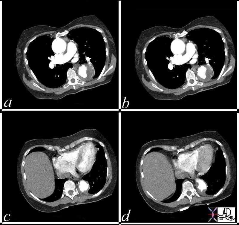
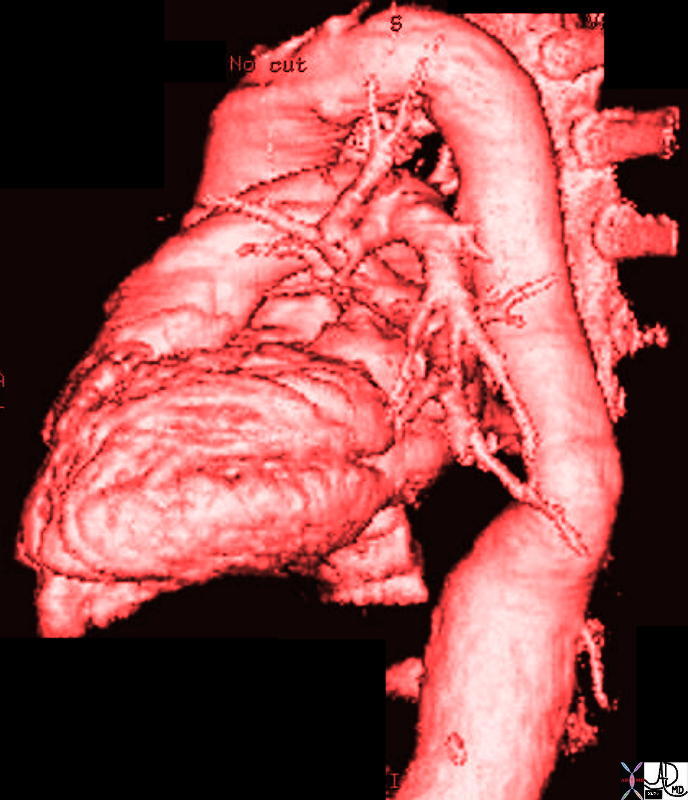
Ascending Aortic Aneurysm |
| This series of images are from a CTscan showing an ascending aortic thoracic aneurysm. There is evidence of heavy calcification of the aortic valve (aortic sclerosis), an aneurysm confined to the ascending aorta (b,c,d), and tortuosity of the descending aorta (d). The cause for the aneurysm is probably a combination of systemic hypertension, aortic stenosis and atherosclerotic degeneration of the wall. Courtesy Ashley Davidoff MD 19426c code CVS thorax aorta ascending aneurysm MAC aortic sclerosis CTscan |
Aortic Arch
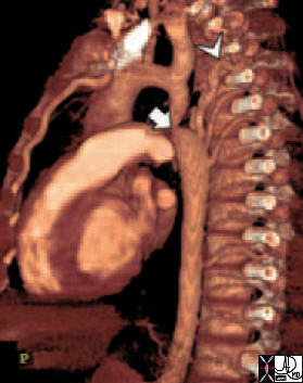
Aortic Arch – Dissection |
| 20449 thorax thoracic aorta arch aorta fx dissection dx aortic dissection type A CTscan Courtesy Ashley Davidoff MD DB |
Contrast CT Masks Thrombosed Dissection Unenhanced Reveals Small Hyperdense Dissection |
| This series of CT scans were taken 1 day apart. The patient presented with chest pain and the soft tissue changes around contrast filled aorta (a,b,c) suggested a chronic dissection, or an acute thrombosed dissection. 1 day later the non-contrast CT clearly reveals an acute thrombosed dissection (d,e,f) that started in the arch and extended along the descending thoracic aorta. The last series (g,h,i) enhance the non contrats study. A non contrast CT followed by contrast injection, is an important tchnique to optimally characterise acute changes in this clincal setting. Courtesy Ashley Davidoff MD 36865c code CVS aorta thoracic arch descending dissection hyperdense cresecent sign acute thrombosed dissection 36865c new |
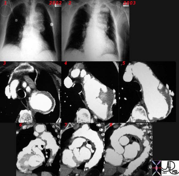
Expanding Aneurysm of the Arch |
| This combination CXR and CT reveals an expanding aneurysm of the arch fromm 2002 to 3003. The CT shows three aneurysm in the arch of the aorta. The largest seen in image 3,6, and 7 accounts for the finding in the left apex of the CXR, while a second pseudoaneurysm is seen on the lateral border of the knob (4,8) and a penetrating ulcer medially (5) 32029c |
Isthmus
 Coartctation – Reconstruction Coartctation – Reconstruction |
| 44146 coarctation of the aorta heart cardiac CTscan MDCT Courtesy Philips Medical |
 Patent Ductus Arteriosus Patent Ductus Arteriosus |
| 42326.800 aorta pulmonary artery finding PDA patent ductus arteriosus left to right shunt arteriovenous malformation CTscan Courtesy Ashley Davidoff MD |
 Ductus Diverticulum Ductus Diverticulum |
| 71175c01 aorta thorax thoracic fx aortic ectasia sigmoid shape tortuosity breast asymmetry size CXR plain film Davidoff MD |
Descending Aorta
Aneurysm |
| This series of CTscans with contrast show a distal thoracic aortic aneurysm showing thrombus in the lumen (a,b) and irregular atherosclerotic plaque. The presence of calcified wall extrinsic to the thrombus makes a fusiform aneurysm likely, and focal thrombosed dissection secondary to ulcer penetration less likely. Courtesy Ashley Davidoff MD 36845c code thorax aorta descending aneurysm thrombus |
Aortic Dissection |
| 20451 descending aorta ascending aorta fx dissection dx aortic dissection type A CTscan Courtesy Ashley Davidoff MD DB |
Abdominal Aorta
Aneurysm |
| 17061.801red aorta chest abdomen aneurysmal disease thoracic aorta abdominal aorta suprarenal aneurysm |
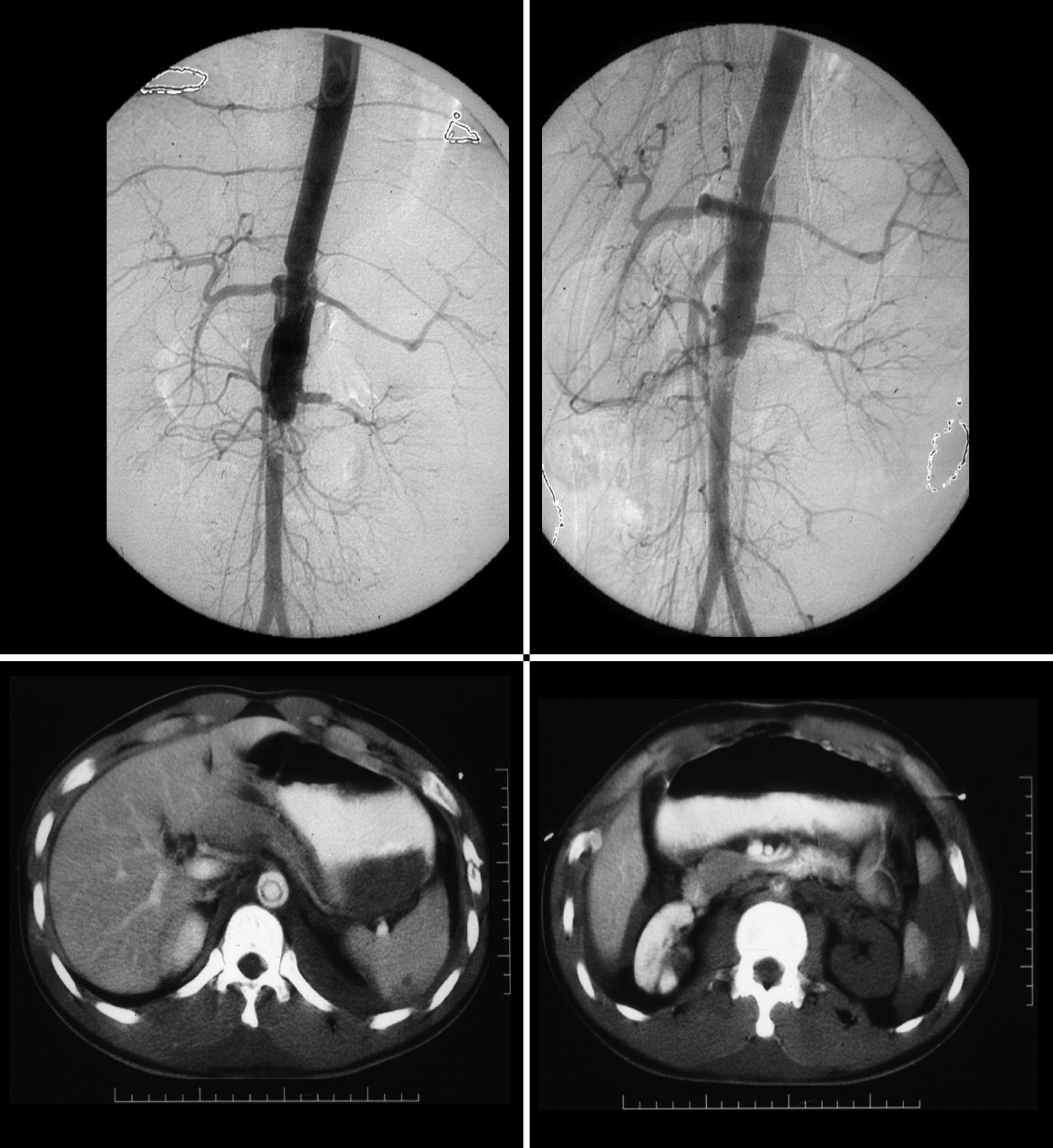
Traumatic Dissection of the Abdominal Aorta wit Loss of Perfusion of the Left Renal Artery |
| These images represent a traumatic dissection of the abdominal aorta caused by a compression injury to the abdomen. The angiograms in a and b shows what appears to be a complex tear of the aorta with a dissection and then a subtotal obstruction of the aorta distally. The CTscan shows (c) the dissection to better effect, with a subtotal narrowing of the aorta distally. Note also the injury of the left renal artery on the angiogram as well as the lack of perfusion of the left kidney on the CT scan. Courtesy Ashley Davidoff MD. 14734c code aorta abdomen trauma dissection |
|
|