
Ultrasound and Echocardiography
Copyright 2007
Subaortic Region
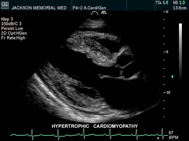
Hypertrophic Obstructive Cardiomyopathy |
| This gray scale echo of the heart shows the left ventricle, anterior and posterior leaflets of the mitral valve, the aortic valve and the base of the aorta. There is a focal thickening of the ventricular septum in the left ventricular outflow tract just proximal to the aortic valve. The region is also slightly more echogenic than the remaining myocardium. This case demonstates a case of asymmetric septal hypertrophy or muscular subaortic stenosis. Courtesy Philips Medical Systems 33134 code cardiac heart echo LV septum thick LVOT narrow echogenic ASH IHSS imaging cardiac echo |
Annulus
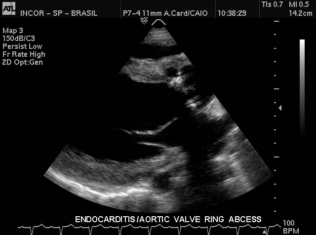
Valve Ring Abscess |
| This gray scale echo of the heart shows the left ventricle, anterior and posterior leaflets of the mitral valve, the aortic valve and the base of the aorta. There is a rounded echogenic focus on the aortic valve ring. In the setting of a febrile illness this represents a ring abscess complicating bacterial endocarditis. Courtesy Philips Medical Systems 33133 code cardiac heart echo MV aortic valve, echogenic, accumulation SBE ring abscess infection imaging cardiac echo |
Aortic Valve
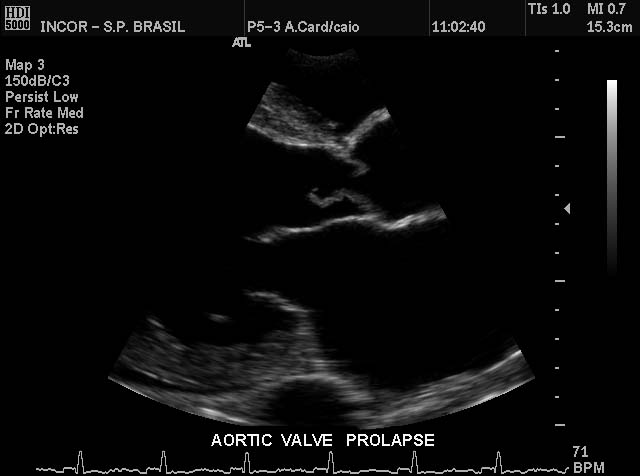
Aortic Valve Prolapse |
| This gray scale echo of the heart showing a right parasternal long-axis left ventricular outflow view, and demonstrating prolapse of the aortic valve. The patient has a diagnosis of aortic regurgitation. Courtesy Philips Medical Systems 33124 cardiac heart echo AOV prolapse MV imaging echo |
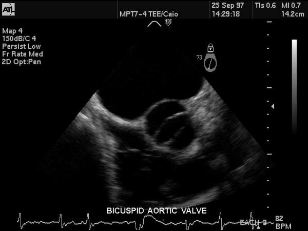
Bicuspid Aortic Valve |
| This gray scale echo of the heart showing a short-axis aorta left atrial view, and demonstrating the aortic valve with two cusps. The patient has a diagnosis of bicuspid aortis valve which is a congenital condition. Courtesy Philips Medical Systems 33169 code cardiac heart echo aorta bicuspid aortic valve congenital imaging cardiac echo |
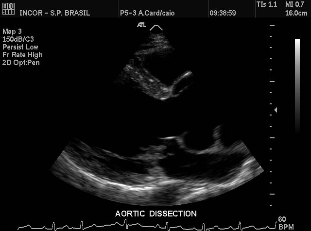
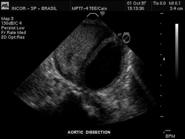
Aortic Dissection |
| This gray scale echo of the heart showing a long-axis left ventricular outflow view, left ventricle, aortic cusps, and base of aorta. Off the far wall of th aorta a dissection originating at the level of the valve is noted. The patient has a diagnosis of aortic dissection. Courtesy Philips Medical Systems 33123 code cardiac heart echo AOV aortic dissection LV AO imaging cardiac echo |
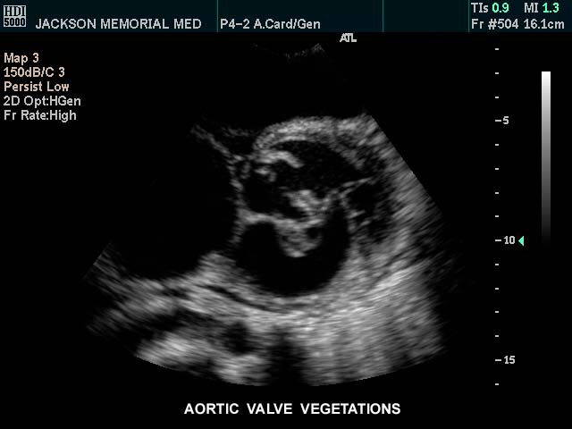
Valve vegetations |
| This gray scale echo of the heart showing a short-axis aorta left atrial view, and demonstrating echogenic vegetations on the aortic valve. The patient has a diagnosis of bacterial endocarditis. Courtesy Philips Medical Systems 33125 code cardiac heart echo AOV vegetation bacterial endocarditis infection imaging cardiac echo |
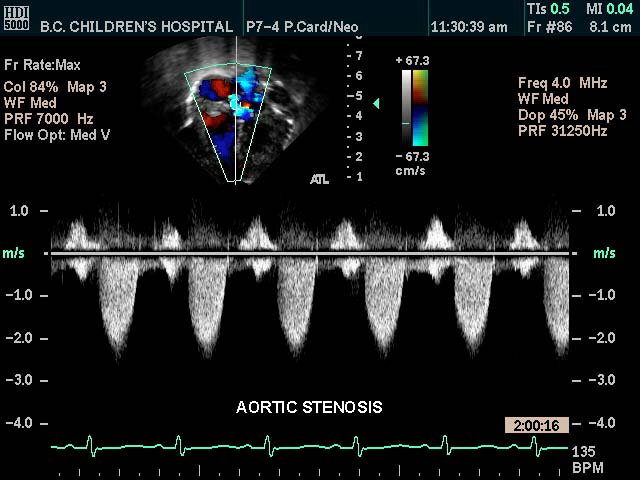
Aortic Stenosis |
| This color flow doppler echo of the heart with pulse flow interrogation at the aortic orifice showing a short-axis aorta left atrial view, and demonstrating high velocity The patient has a diagnosis of aortic stenosis Courtesy Philips Medical Systems 33144 code cardiac heart echo aorta AOV AV AS imaging cardiac echo tube principle |
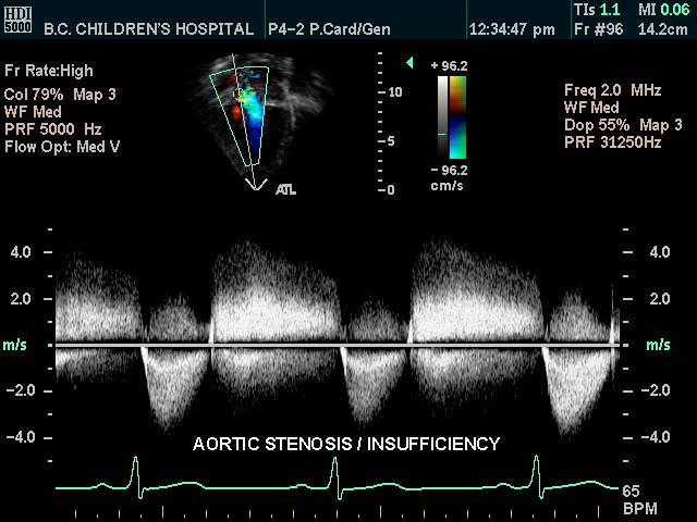
Aortic Stenosis and Insufficiency |
| This color flow doppler echo of the heart with pulse flow interrogation of the aortic valve showing high velocity flow both antegrade and retrograde across the valve indicating both aortic stenosis and aortic regurgitation. Courtesy Philips Medical Systems 33145 code cardiac heart echo pulsed doppler color aorta valve AR AS AOV imaging cardiac echo |
Sinuses
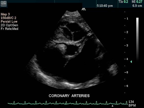
Normal Coronary Artery Arising off the Right Coronary Sinus |
| This gray scale echo of the heart showing a short-axis aorta left atrial view, and demonstrating the coronary arteries and a normal tricuspid pulmonary valve. The right coronary artery is particularly well seen. Courtesy Philips Medical Systems 33146 code normal cardiac heart echo coronary artery pulmonary valve imaging cardiac echo |
|
Normal Coronary Artery Arising off the Right Coronary Sinus |
| This gray scale echo of the heart showing a short-axis aorta left atrial view, and demonstrating the coronary arteries and a normal tricuspid pulmonary valve. The right coronary artery is particularly well seen. Courtesy Philips Medical Systems 33146 code normal cardiac heart echo coronary artery pulmonary valve imaging cardiac echo |
Ascending Aorta
Aortic Arch
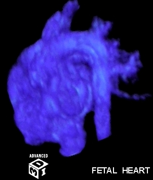
Normal Fetal Heart |
| This image is a 3D reconstruction of a fetal heart in sagittal view, showing the aorta with brachiocephalic vessels, the left atrium abutting the descending aorta posteriorly, and the right ventricle situated anteriorly. No other technology has such an exquisite ability to image the fetal heart. Courtesy of Philips Medical Systems, Ultrasound 32143 code heart normal fetus imaging cardiac echo overlay |
Aortic Knob
Isthmus
Descending Aorta
|
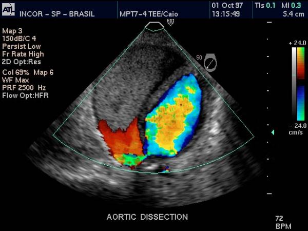
Dissection of Descending Aorta |
| Doppler US of the descending thoracic aorta showing flow in the true lumen (color) and no flow in the thrombosed lumen (gray echoes) in this patient with aortic dissection. Courtesy Philips Medical Systems 33166 |
Abdominal Aorta