|
Plain X-Ray
The Common Vein Copyright 2007
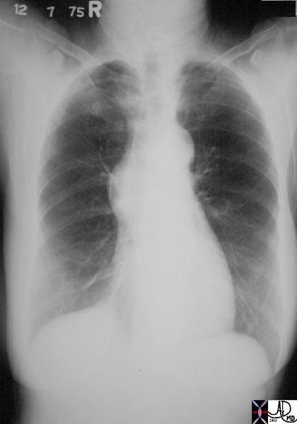
Annulus
Calcified Aortic Annulus
|
| 72864.801c01 72864.800 aorta aortic annulus fx calcified calcification dx aortic sclerosis CXR plain film of chest Courtesy Ashley DAvidoff MD |
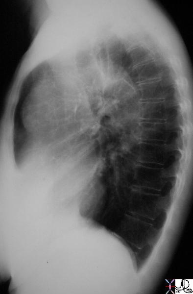
Aortic Valve

Calcific Aortic Stenosis and Sclerosis
|
| 47368c02 spine bone thoracic spine ospeopenia osteoporosis fx wedge compression fractures shape mechanical forces calcification aortic valve aortic sclerosis aortic stenosis AS kyphosis CXR plain film X-ray Davidoff MD |
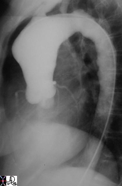
Ascending Aorta
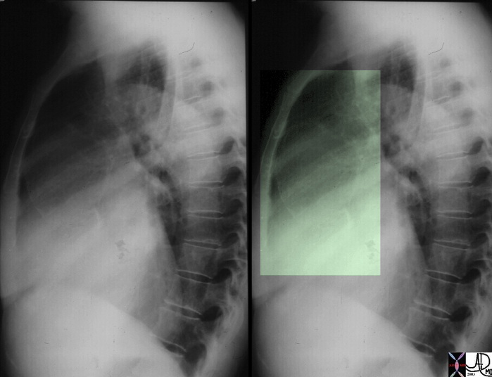
Tertiary Syhpilis Ascending Aorta – Dilated and Calcified
|
| This lateral examination of the chest shows fine calcification in an ectatic ascending aorta associated with aortic annular calcification. Note the remarkable paucity of atherosclerotic change in the descending aorta. These findings are highly characteristic of tertiary syphilis of the aorta. Courtesy Ashley Davidoff MD. 00018c code CVS artery aorta ascending syphilis aneurysm calcification tortoise shell |
Aortic Arch
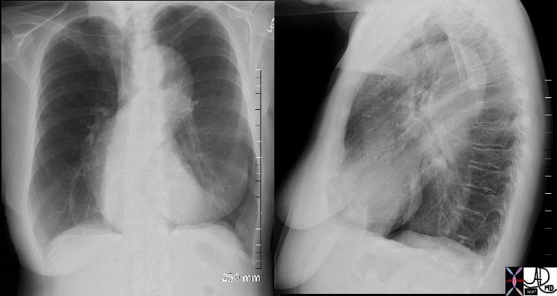
Aortic Arch – Pseudoaneurysm
|
| This image represents a combination of plain film CXR and the correlative thoracic aortogram in a 38 year old patient, 13 years after an MVA. There is an aneurysmal bulge at the level of the isthmus, representing a traumatic aneurysm at the characteristic location of the ligamentum arteriosum. The P-A and lateral chest X-ray shows an enlarged and unusually shaped aortic knob and the angiogram confirms the pseudoaneurysm of the aorta. 35178c Courtesy of Laura Feldman MD. code CVS aorta artery thorax trauma |
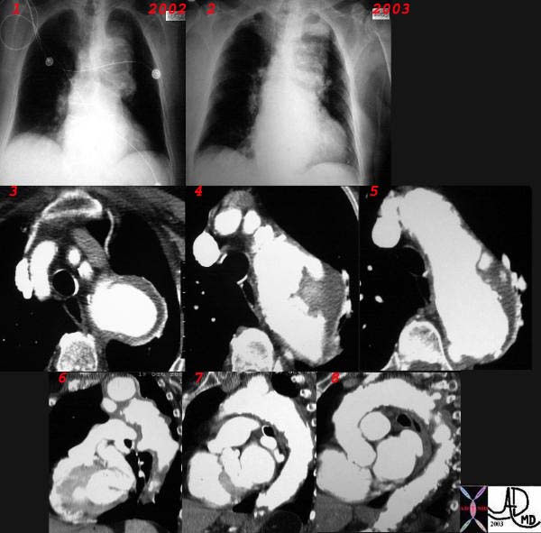
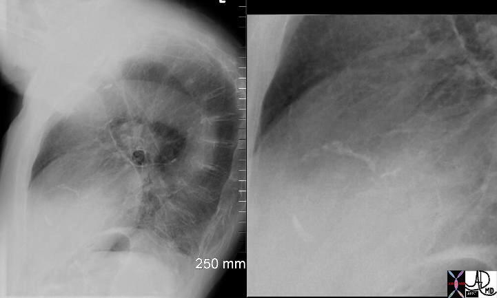
Expanding Aneurysm of the Arch
|
| This combination CXR and CT reveals an expanding aneurysm of the arch fromm 2002 to 3003. The CT shows three aneurysm in the arch of the aorta. The largest seen in image 3,6, and 7 accounts for the finding in the left apex of the CXR, while a second pseudoaneurysm is seen on the lateral border of the knob (4,8) and a penetrating ulcer medially (5) 32029c |
Aortic Knob
Calcified Mass
|
| 46008c01 chest aorta mediastinum fx calcified mass with rim calcification post op clips fx dystrophic calcification shape character CXR plain X-ray of chest CTscan Davidoff MD 46008.800 46008c01 |
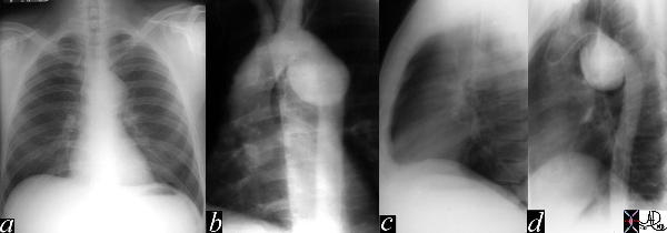
Isthmus
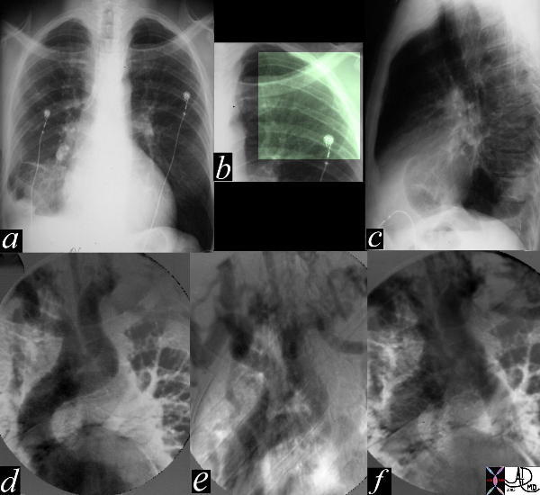
Aortic Interruption – Severe Coarctation
|
| The most obvious finding in this CXR (a) with pleuro-parenchymal changes is not the most significant. In image (b) the highlighted ribs reveal rib notching characteristic of coarctation of the aorta. The lateral examination (c) in this instance is not helpful. In the early phase of the angiogram(d), there appears to be complete interruption of the aorta with a large left subclavian artery acting as a collateral pathway. The sbsequent images e, and f, show progressive filling of the isthmus and distal thoracic aorta. The coarcatation becomes apparent characterised by a “3” sign. 35107c Courtesy Laura Feldman MD code CVS artery aorta thorax coarctation rib notching bone collateral |
Descending Aorta
Tortuous Descending Aorta
|
| 71175c01 aorta thorax thoracic fx aortic ectasia sigmoid shape tortuosity breast asymmetry size CXR plain film Davidoff MD |
Abdominal Aorta
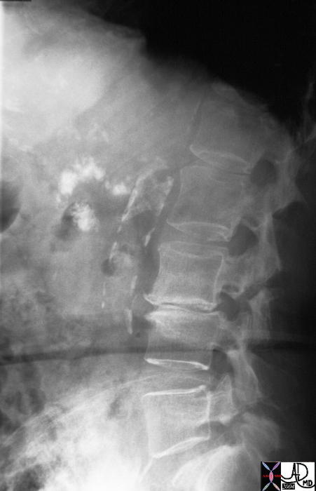
Calcified Atherosclerosis
|
| 32916 Courtesy Ashley Davidoff MD abdomen calcification pancreas calcified body tail uncinate process chronic pancreatitis dystrophic calcification medical students inflammation imaging radiology lateral abdomen aorta radiologists and detectives |
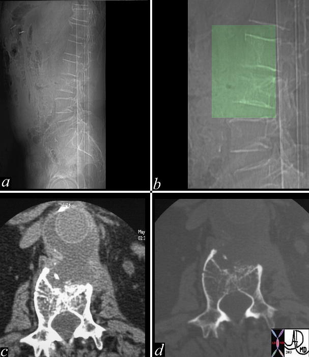
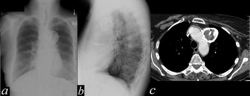
Scalloping of Vertebra and Mycotic Aneurysm
|
| This combination of images from a CTscan of the abdomen are of a middle aged man who presented with back pain and fever, with a remote history of AAA repair. The lateral scout film shows scalloping of vertebral bodies 2 and 3 (a) highighted in green overlay in b. The CTscan with soft tissue windows (c) and bone windows (d) show a complex fluid collection surrounding the aorta which proved to be a perigraft infection. Courtesy Ashley Davidoff MD. 22725c02 code CVS artery aorta abdomen abscess AA repair infection bone vertebra lumbar anterior scalloping |
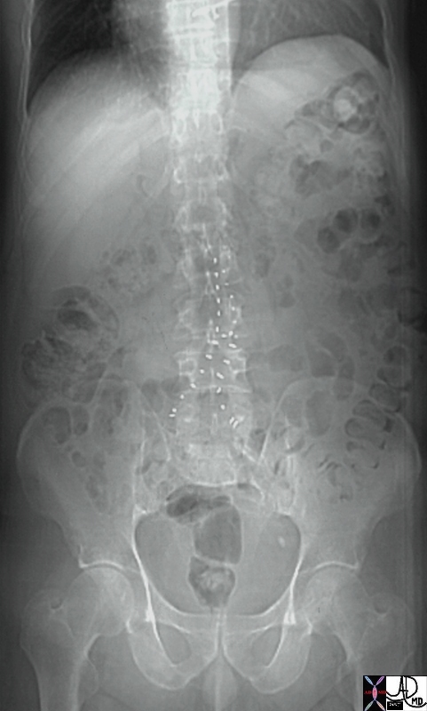
Stent Graft
|
| 24284b01 aorta AAA repair abdominal aortic aneurysm MIT stent graft X-ray plain film KUB Davidoff MD |
|