
Copyright 2007
Ashley Davidoff MD
Introduction
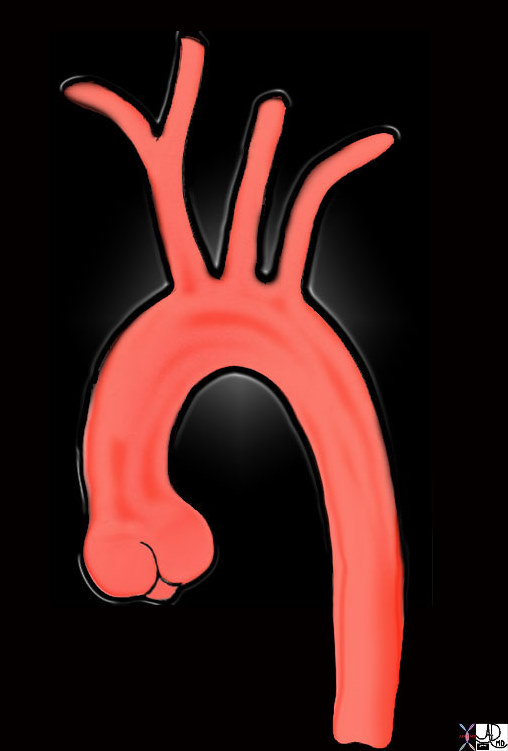
The aortic arch is the segment between the ascending and descending thoracic aorta. Its course begins at the upper level of the second right sternocostal junction. The arch runs upwards in an anterior-superior curve towards the left in front of the trachea. It then proceeds downward passing the left side of the body of the fourth thoracic vertebra and then meets the proximal descending aorta. The aortic arch is characterized structurally by its tubular curvature and elastic properties. At the top of the arch are three arterial branches called as a group the brochiocephalic arteries which carry blood to the head and upper limbs. Common diseases include atherosclerosis and aortic aneurysm. Dissection commonly involves the arch, often originating just beyond the left subclavian artery. Diagnostic studies include CT scanning, aortography, MRI and echocardiography. Treatment is usually surgical.
keywords aorta aortic valve sinotubular junction ascending aorta aortic arch brachiocephalic artery left common carotid artery left subclavian artery descending aorta normal TCV Ashley Davidoff MD TheCommonVein.net 47676
The aortic arch begins where the ascending aorta leaves the pericardium, at or near the origin of the first brachiocephalic artery and ends after the origin of the last brachiocephalic artery, (usually left subclavian) at the level of the ligamentum arteriosum.
The three branches of the arch are the brachiocephalic artery, left common carotid and left subclavian artery. The brachiocephalic artery gives rise to the right subclavian and right common carotid arteries. There are normal variations of this pattern. The most common variation is when the brachiocephalic artery and the left common carotid have a common origin. Sometimes the vertebral arteries arise as independant branches off the arch. The aberrant right subclavian artery is one of the other common variations named when the right subclavian artery arises from the left side as the last branch of the arch, after the left subclavian artery. It thus has to make its way from the left side to the right by crossing behind the trachea and esophagus.
There are three branches that extend from the aortic arch. The innominate artery, (which quicky branches again into the right common carotid and right subclavian arteries), the left common carotid artery, and the left subclavian artery.
There are numerous variations in just how many branches originate from the arch. In rare cases, the left carotid and left subclavian can originate from the innominate, making the innominate the only branch extending from the arch. On the other extreme, there have been cases where 5 or 6 branches extend from the arch. In these cases, the internal and external carotids arise directy from the arch (and therefore, the common carotid is absent).
Parts
Consists of the arch itself and the isthmus. The aortic isthmus, a narrowing of the vessel, is present betwene the left subclavian artery’s and the attachment of the ligamentum arteriosum, a remnant of the ductus arteriosus from fetal development, in adults present as a fibrus cord attaching the arch of the aorta to the left pulmonary artery. Beyond the ligamentum arteriosum is a spindle-shaped dilation called the aortic spindle. The branches of the arch of the arta are the braciocephalic trunk, the left common carotid artery, and the left subclavian artery, which arise from the arch’s highest point. There are a number of congenital variations in which the vessels can arise and branch
Size
2.5-3cms need to check this
Shape
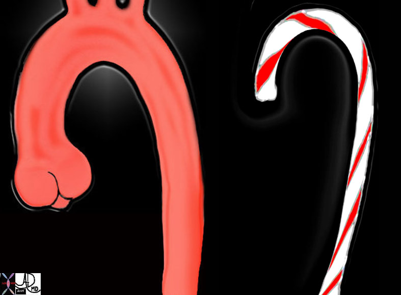
The arch starts just anterior to the trachea in a relatively rightward position and forms two arches. The first is the more easily undestood vertical arch formed as it ascends up and over the left mainstem bronchus. It is often described as being candy cane in shape.
keywords candy cane shape aorta aortic valve sinotubular junction ascending aorta aortic arch descending aorta normal
Ashley Davidoff MD TheCommonVein.net 47677c03
However when viewed in the frontal projection the arch is not qiute perfect.
Angle of the Arch in this Projection has an Acute Component |
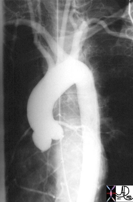
| 27185 This angiogram of the normal thoracic aorta is taken in the LAO projection. The thoracic aorta consists of a bulbous portion called the aortic sinus that is the most proximal portion, and is connected to the tubular portion that connects the sinus portion to the aortic arch. The isthmus connects the aortic arch to the descending thoracic aorta.
(see 27185g for overlay image) Courtesy Ashley Davidoff MD 27185 code CVS aorta thorax ascending sinus sinotubular junction tubular AOV arch isthmus descending |
Note that the angle between the arch and descending aorta is too acute.
This aberrancy is due to the a second bed or arch in the aorta as it crosses the the trachea. The arch of the aorta thus forms two curves, one with covexity upward and over the trachea and the second an almost horizontal bend around the trachea first first to the left and then posterior.
Images courtesy of: Ashley Davidoff, M.D. TheCommonVein.net
aorta Philips 026
Thus when you view the arch of a conventional thoracic aortogram, you will be able to understand why it does not look like a perfect vertical arch – it is also bent slightly folded around the trachea and esophagus. In the elderly the aortic knob becomes prominent because of unfolding of the aorta. This means that the second bend flattens making the knob more prominent.

This angiogram of the normal thoracic aorta is taken in the left anterior oblique (LAO) projection. In this projection the shape is not quite a candy cane suggesting some complexity to the arch and known when seen as a three-dimensional structure. Image courtesy of: Ashley Davidoff, M.D. TheCommonVein.net aorta Philips 025
Position and Relations
Its origin is level with the upper border of the sternocostal articulation of the right side; from there it courses upward and left, in front of the trachea, then backwards to the left of the trachea, and finally downward to the left of the fourth thoracic vertebra, at the bottom of which it becomes the descending aorta. The artery usally rises to a height of 2.5 cm below the upper border of the manubrium sterni. In some patients, the arch of the aorta may rise to the top of the sternum instead of 2.5 cm below its top. In other patients the distance downard from the top is 4 cm, and more rarely, 5-8 cm.
On the arch of the aorta’s left side in this portion are four nerves: the left phrenic nerve, the caudal of the superior cardiac branches of the left vagus nerve, the superior cardiac branch of the left sympathetic nerve, and the trunk of the left vagus nerve, the last of which gives off a recurrent branch that hooks around the arch of the aorta and continues upward on the artery’s right side. Between the phrenic and vagus nerves, the highest left intercostal vein courses to the arch of the aorta’s left side. On the vessel’s right there is the deep part of the cardiac plexus, as well as the right recurrent nerve, the esophagus, the thoracic duct, while in back and to the right is the trachea. Above it are its own three branches, the brachiocephalic trunk, the left common carotid artery, and the left subclavian artery, to whose origins the brachiocephalic vein comes close. Below the arch of the aorta there is the bifurcation of the pulmonary artery, as well as the left bronchus, the superficial part of the cardiac plexus, and the left recurrent nerve, and the ligamentum arteriosum, a a remnant of the ductus arteriosus from fetal development, in adults present as a fibrous cord attaching the arch of the aorta to the left pulmonary artery.
Occasionally the arch of the aorta runs over the right mainstem bronchus and not the left, and passes along the vertebral column’s right side. This is called a right aortic arch. In such a case the abdominal viscera may also be transposed. A double aortic arch may also occur due to the duel origin of the vessel..
Covering
The arch of the aorta is covered in front by the lungs’ anterior margins, the pleura, and the remains of the thymus; the left side of it, while running backward, touches the left lung and pleura.
Applied Anatomy
Sometimes the aorta arches over the root of the right lung (right aortic arch) instead of over that of the left, and passes down on the right side of the vertebral column. In such cases all the thoracic and abdominal viscera are transposed. Less frequently the aorta, after arching over the root of the right lung, is directed to its usual position on the left side of the vertebral column; this peculiarity is not accompanied by transposition of the viscera.
Size Changes
Aneurysms of the Arch
Can use defn of aneuysm already describe Cause athero result progressive dil with risk of Rupture DX CT Echo MRI aortography Rx surgical based on size
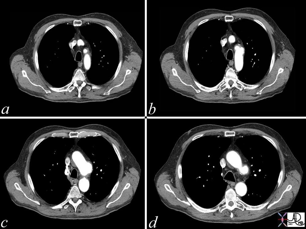
Focal Aneurysm of the Arch of the Aorta |
| A series of CT scans showing a focal aneurysm of the aortic arch orignating just beyond the left subclavian artery. Whether this represents a focal saccular aneurysm from focal atherosclerotic weakening, or an ulcerating and penetrating plaque, or a site of previous dissection is difficult to know. Courtesy Ashley Davidoff MD. 36817c code CVS chest thorax aneurysm aorta arch |
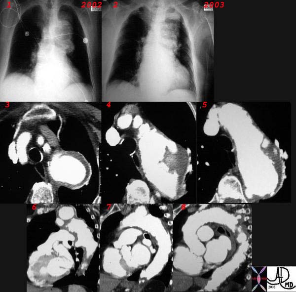
Focal Aneurysm of the Arch |
| This combination CXR and CT reveals an expanding aneurysm of the arch fromm 2002 to 3003. The CT shows three aneurysm in the arch of the aorta. The largest seen in image 3,6, and 7 accounts for the finding in the left apex of the CXR, while a second pseudoaneurysm is seen on the lateral border of the knob (4,8) and a penetrating ulcer medially (5) 32029c |
Hypoplastic Arch
Can use defn of HLHS (hypoplastic left heart syndrome) already describe Cause congenital result progressive poor output to systemic circ particulalrly to brain DX Echo MRI Rx surgical if possible Norwood if part of more complex HLHS
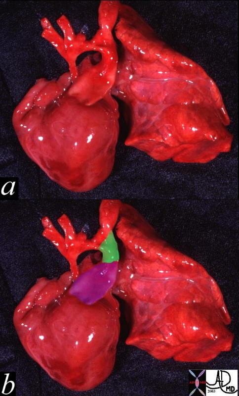
Hypoplastic Arch and Ascending Aorta |
| This pathologic specimen shows a small ascending aorta, and arch with a diffuse coarctation. with a large pulmonary artery (purple overlay in b) and a patent ductus arteriosus (green overlay in b)Courtesy Ashley Davidoff MD 00264c code heart cardiac hypoplastic aortic arch PDA coarctation of the aorta AO congenital grosspathology |
Right Aortic Arch
defn Cause congenital may be isolated maybe in conjunction with other CHD (congenital heart disease) or other visceral abnormalities Associated particulalrly with Tetralogy of Fallot and truncus arteriosus result as an anomaly has no significance but associated diseases have functional significance DX CXR Echo MRI Rx not indicated but directed to searchand Rx associated anomalies
Right Aortic Arch |
| 35334 Courtesy of Laura Feldman MD. code aorta arch artery right aortic arch thorax |
Cervical arch
defn Cause congenital may be isolated maybe in conjunction with other CHD (congenital heart disease) Associated particularly with pseudo-coarctation result as an anomaly has no significance DX CXR MRI Rx not indicated
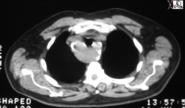
Pseudcoarctation |
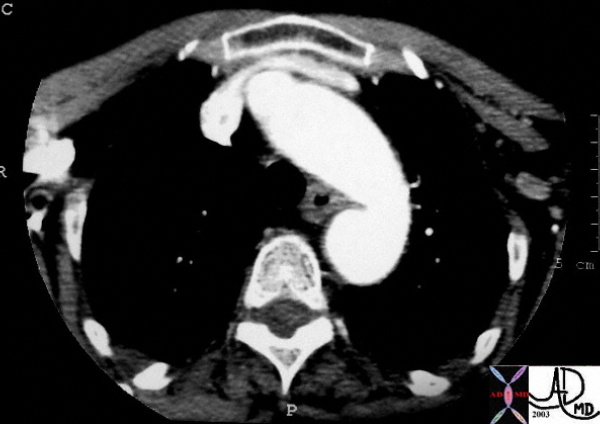
| This cross sectional view of the chest at the apex of the lungs at the level of the manubrial notch shows the top of the aortic ach which is more superior than usual and is known as a cervical arch. The aorta more inferiorly is also redundant and tends to kink eventually appearing as a pseudocoarctation. In this case only a kink is present. Courtesy Ashley Davidoff MD 33539 see 33535 code aorta arch cervical kink pseudocoarctation imaging radiology CTscan |
Pseudocoarctation
defn Cause congenital may be isolated maybe in conjunction with cervical arch result as an anomaly has no significance DX CXR MRI cathterisation to identify and eval for pressure gradient Rx not indicated
Pseudocoarctation |
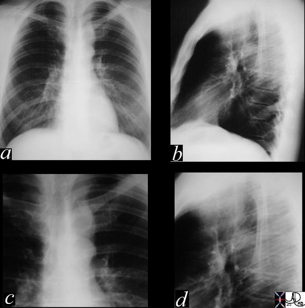
| This series of CXRays P-A (a) lateral (b) and magnified views (c,d) showing an aberrant shape to the aortic knob which is characterised by a “3 sign” reminiscent of a coarctation segment. There is no assocated rib notching nor a pressure gradient and hence the diagnosis is a pseudocoarctation of the thoracic aorta. 35187c01 Courtesy LAura FEldman MD code CVSartery aorta thorax 3 sign peudocoarctation. |
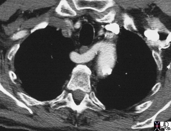
Aberrant Right Subclavian Artery |
| 16376 aorta aberrant origin of right subclavian artery as the last vessel off the left aortic arch congenital growth position esophagus CTscan Davidoff MD |
Dissection
Stick and paste previous def from ascending aorta
specific issue is the involvement of the arteries to head and potential for a stroke
Aortic Dissection |
| 20449 thorax thoracic aorta arch aorta fx dissection dx aortic dissection type A CTscan Courtesy Ashley Davidoff MD DB |
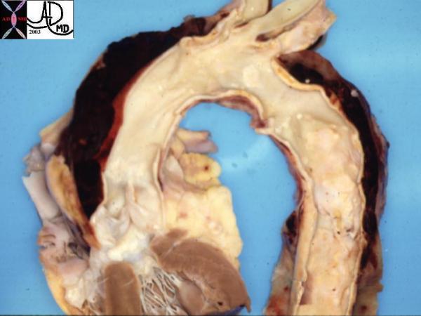
Aortic Dissection Type A |
| This pathological specimen shows an aortic dissection starting at the root of the aorta and extending across the arch and into the descending portion. The false lumen is filled with clotted blood. Courtesy Henri Cuenoud MD 13421 code CVS thorax AO aorta dissection grosspathology |